The brain’s wiring network could help predict how aggressive gliomas are
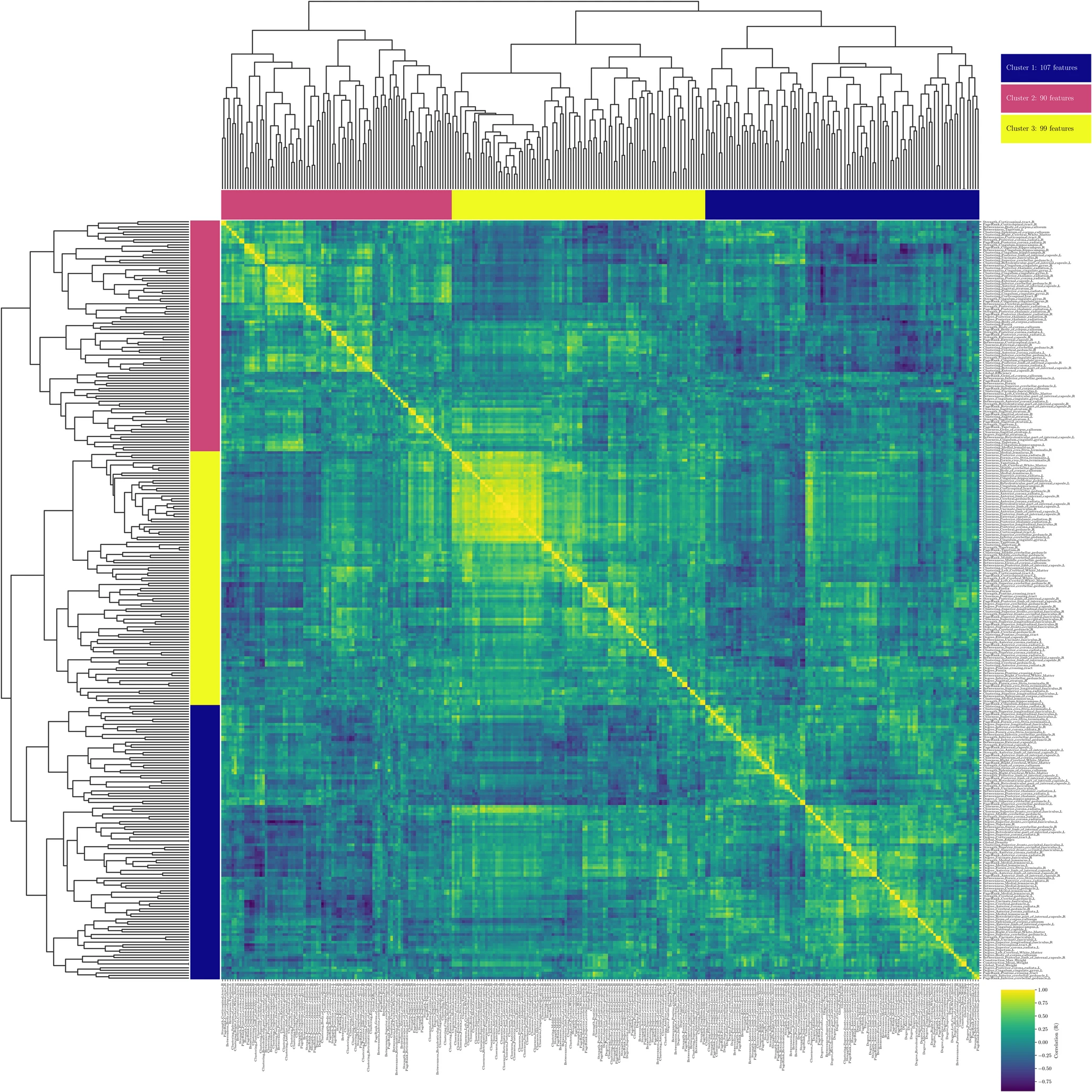
A pilot study published in Neuroradiology, with the participation of iHEALTH researchers, combines clinical MRI and interpretable artificial intelligence to distinguish low-grade from high-grade gliomas based on how they disrupt the brain’s white matter.
Gliomas are the most common primary brain tumors, but they don’t all behave the same way. Some grow slowly over years; others progress rapidly. Knowing before surgery whether a glioma is low- or high-grade helps the neuro-oncology team decide how much tissue to remove without putting functions such as language or movement at risk. It also helps them estimate the prognosis and plan follow-up treatment.
The definitive diagnosis comes from analyzing the tissue obtained through biopsy, which today is complemented by molecular testing. Before that stage, conventional MRI shows the tumor’s shape, but it doesn’t capture well the impact the lesion has on the rest of the brain. That is the problem addressed by a pilot study published in the journal Neuroradiology. The authors include iHEALTH researchers Steren Chabert and Rodrigo Salas, from Universidad de Valparaíso, together with Cristian Montalba, Ignacio Espinoza and M. Daniela Cornejo, from Pontificia Universidad Católica de Chile; Pamela Franco, from Universidad Andrés Bello; and Francisco Torres and Carlos Bennett, from Hospital Carlos Van Buren.
The work is based on the connectome, the map of the brain’s connections. “Imagine the brain as a large highway network. The connectome is the map of that network: it shows which areas are connected to each other and how much information ‘traffic’ flows between them,” the researchers explain.
To build it, the team analyzed 50 white matter tracts, the fiber bundles that link different brain regions, reconstructed for each patient using diffusion MRI. From this perspective, a glioma is no longer just an isolated lesion. “A tumor can cut off main routes, force detours, and leave areas far away from it less well connected,” they note.
A key aspect of the method is what is known as tumor masking: excluding the areas occupied by the tumor from the analysis. Within the lesion, tissue deformation and inflammation produce false connections that contaminate the calculations. “By masking the tumor, we measure the real impact on the remaining network rather than artifacts within the tumor mass,” they explain. The hypothesis is that a high-grade glioma, being more infiltrative, disrupts the surrounding white matter far more.
The study included 35 patients with confirmed glioma (20 low-grade and 15 high-grade) who underwent surgery at a single center. Their images were acquired before surgery on a standard 1.5 Tesla clinical scanner. This was a deliberate choice, made to test whether the method works with the equipment used in routine practice.
An artificial intelligence that explains its decisions
Using each patient’s connectome data, the team trained a machine learning model that correctly classified 82.9% of patients. This is a preliminary estimate obtained within the same cohort. When clinical variables such as tumor volume and location were added, the estimated performance improved, although the authors caution that this difference has not yet been demonstrated.
Beyond the figure, the focus is on the model being interpretable. “A ‘black box’ model delivers a binary verdict, ‘high-grade’ or ‘low-grade’, without explaining how it reached that conclusion,” they explain. Using a tool called SHAP, the team was able to see which tracts carried the most weight in each decision. The most informative were the sagittal stratum, the medial lemniscus and the superior fronto-occipital fasciculus: high-grade tumors were associated with a loss of centrality and connection strength in these long pathways.
“For the medical team to understand the model’s reasoning is essential to verify whether it makes anatomical sense,” the researchers state. “No one should make a surgical decision based on a number they can’t question.”
The authors stress that this is a feasibility study. “It’s still a long way off, and we want to be very clear on this point. This work is a pilot feasibility study, not a tool ready for the operating room,” they say. To reach the clinic, the approach must be validated across multiple hospitals and with different MRI scanners. It should always complement conventional MRI and pathology, never replace them.
Next steps include analyzing larger, multicenter cohorts and incorporating molecular biomarkers to align the findings with the current WHO classification. “The big scientific question is whether these connectivity signatures correlate with the tumor’s genetic subtypes, or whether they provide independent prognostic information that genetics doesn’t capture,” they say. Looking ahead, they also aim to integrate other techniques, such as perfusion MRI.
Franco, P., Montalba, C., Espinoza, I. et al. Tumor-masked structural connectomics for assessing tract-specific white matter disruption associated with glioma aggressiveness: an interpretable machine learning pilot study. Neuroradiology (2026). https://doi.org/10.1007/s00234-026-04232-0
