Successful First Stream 1 Meeting of the Year
This meeting, held every semester, aims to share the latest results from iHEALTH and expose master's and doctoral students to cutting-edge research in medical imaging. It is an opportunity that strengthens the institute's scientific community and shapes its work for the future.
On Thursday, April 9, the San Agustín Auditorium at the Catholic University of Chile hosted a new Stream 1 (S1) Meeting, led by principal investigator René Botnar. S1 is one of the four interdisciplinary working groups within the Institute and aims to develop methods based on physics and artificial intelligence to optimize scan times, expand quantifiable parameters, and improve disease diagnosis.
"Overcoming the current limitations of medical imaging, such as the lack of comprehensive quantification, integration with other clinical data, and high operating costs, through the development of innovative techniques that integrate AI, image physics, and multiparametric data, from acquisition to diagnosis," as stated in the group's description.
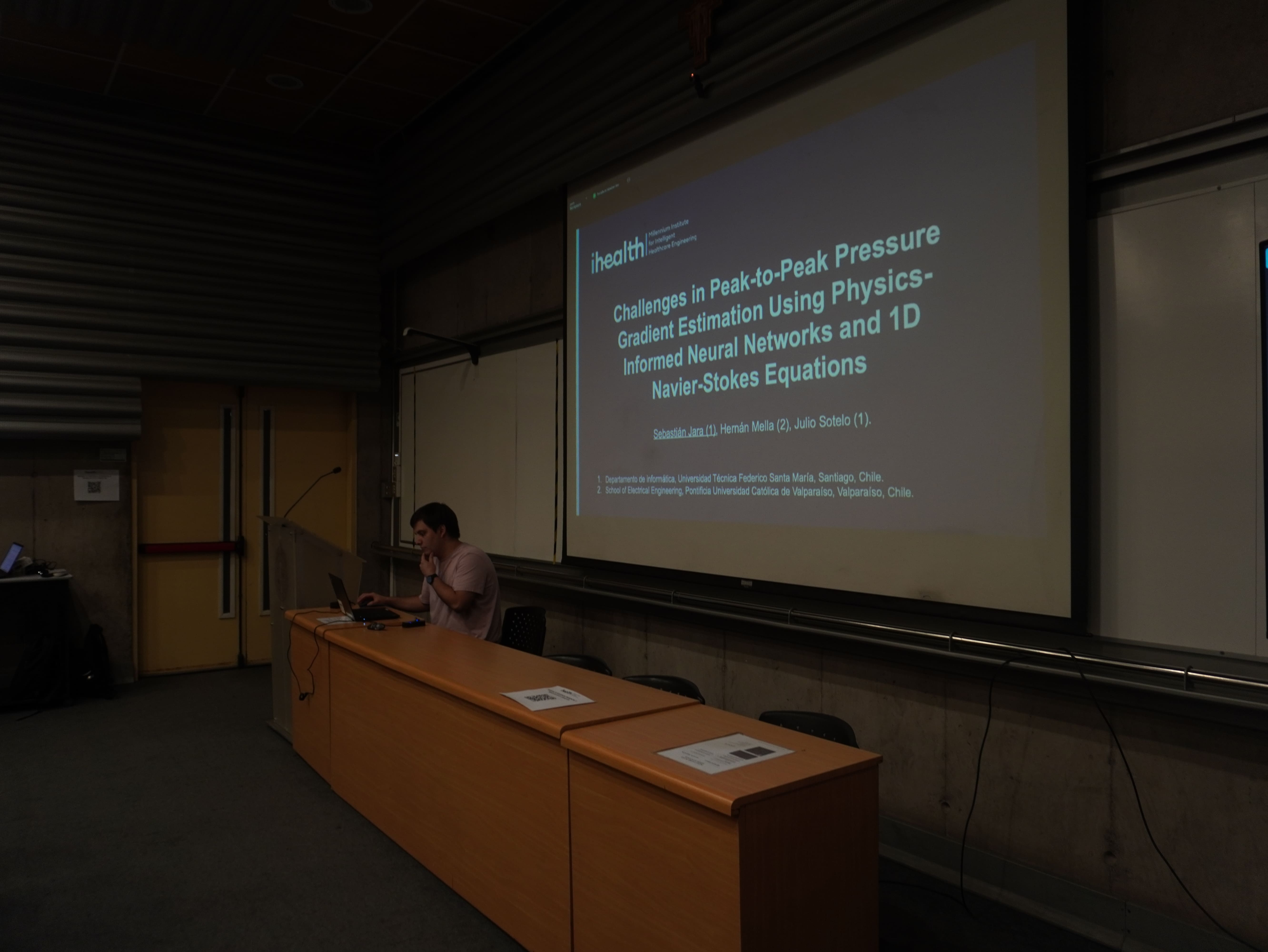
The event began with a remote presentation by doctoral student Sebastián Jara, who detailed his work on the use of neural networks to non-invasively estimate blood vessel pressure, a parameter that typically requires catheter-based measurement.
"The use of artificial intelligence models based on Physics-Informed Neural Networks (PINNs), integrating reduced 1D models of the Navier-Stokes equations, allows for the non-invasive estimation of the pressure gradient in patients with aortic coarctation," the researcher explained. These models have low computational cost, making them particularly suitable for future application in clinical settings.
She added, "The results in patients with aortic coarctation show good agreement with measurements obtained by catheterization, demonstrating the potential of these techniques to support cardiovascular diagnosis using magnetic resonance imaging data."
Next, Dabne Barrera presented the results of her research, which develops and compares two new 3D protocols for simultaneous T1-T2 mapping of hyaline articular cartilage in the knee at 0.55T. This mapping allows for the characterization of cartilage in patients with osteoarthritis, providing information on fibrosis, inflammation, and edema.
The researcher evaluated the accuracy, precision, and sharpness of the maps, as well as the reconstruction time. "Both protocols achieved complete knee coverage with isotropic resolution of 1 mm in 3:48 min (Cartesian) and 3:40 min (radial). The proposed protocols allow for rapid and isotropic quantification of knee cartilage at 0.55T in approximately 4 minutes, with accurate, precise, and repeatable measurements," he emphasized.
The meeting concluded with research professional Pablo Pino, who presented advancements in low-field magnetic resonance imaging (MRI) of the lungs, specifically in the development of a 3D free-breathing sequence that allows for ventilation estimation and the diagnosis of conditions such as pulmonary fibrosis.
"Computed tomography (CT) remains the most widely used modality for pulmonary evaluation; however, repeated exposure to ionizing radiation carries risks that limit its use in serial studies and long-term monitoring," he noted.
The project Pablo is developing is designed to overcome conventional limitations and has the potential to reduce costs and infrastructure requirements. "Our approach integrates advanced motion correction with free-breathing acquisitions, without the need for apnea. Validation in healthy volunteers demonstrated the feasibility and robustness of this technique; in all respiratory phases, the pulmonary vasculature and other structures were clearly delineated with consistent image quality," he concluded.