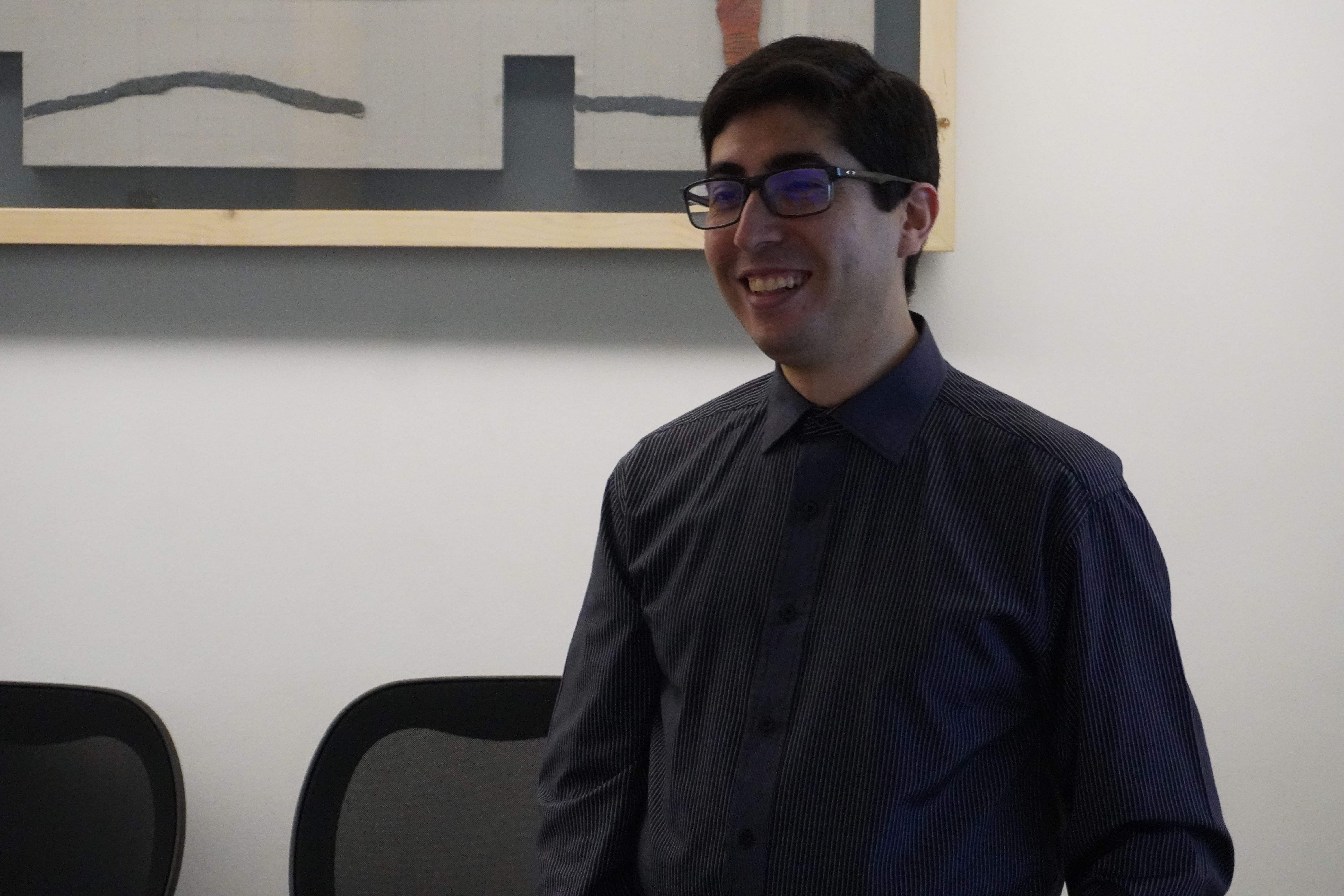
Pablo Messina successfully defends doctoral thesis on more reliable AI-generated radiology reports
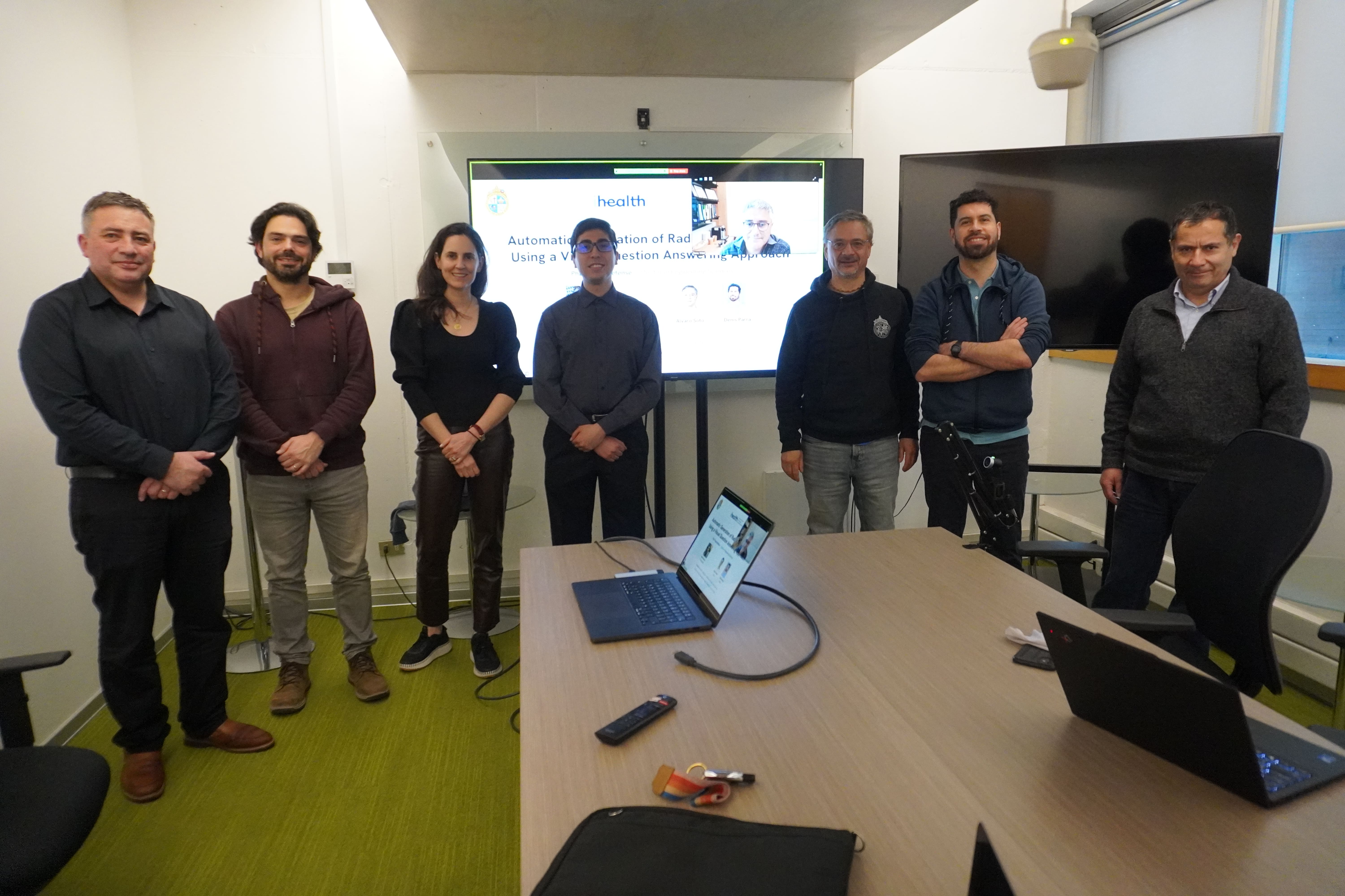
The researcher earned his PhD in Engineering Sciences from Pontificia Universidad Católica de Chile with a thesis that proposes generating chest X-ray reports by answering specific clinical questions about the image instead of writing free text. His work culminated in an oral presentation at CVPR 2026, the world’s leading computer vision conference.
The chest X-ray is the most frequently requested imaging study in clinical practice, and each one requires a report that guides medical decisions. “The report is the bridge between the image and the clinical decision,” Messina explains. “But writing it requires a specialist with years of training and time for each exam, and in high-demand hospitals, exams pile up faster than they can be reported.”
Artificial intelligence can help, but current models produce text that sounds correct and still misses findings, confuses negations, or invents abnormalities. They also rarely show which part of the image they rely on. Messina’s thesis tackles these problems by breaking the report down into specific clinical questions. Some can be answered with yes or no, such as “Is there a pleural effusion?” Others ask the model to locate and describe an anatomical region, such as the right clavicle. “If the report is built from answers to specific questions, each statement can be verified separately and is linked to a region of the image. That way, if there’s an error, you can see where it is,” he notes.
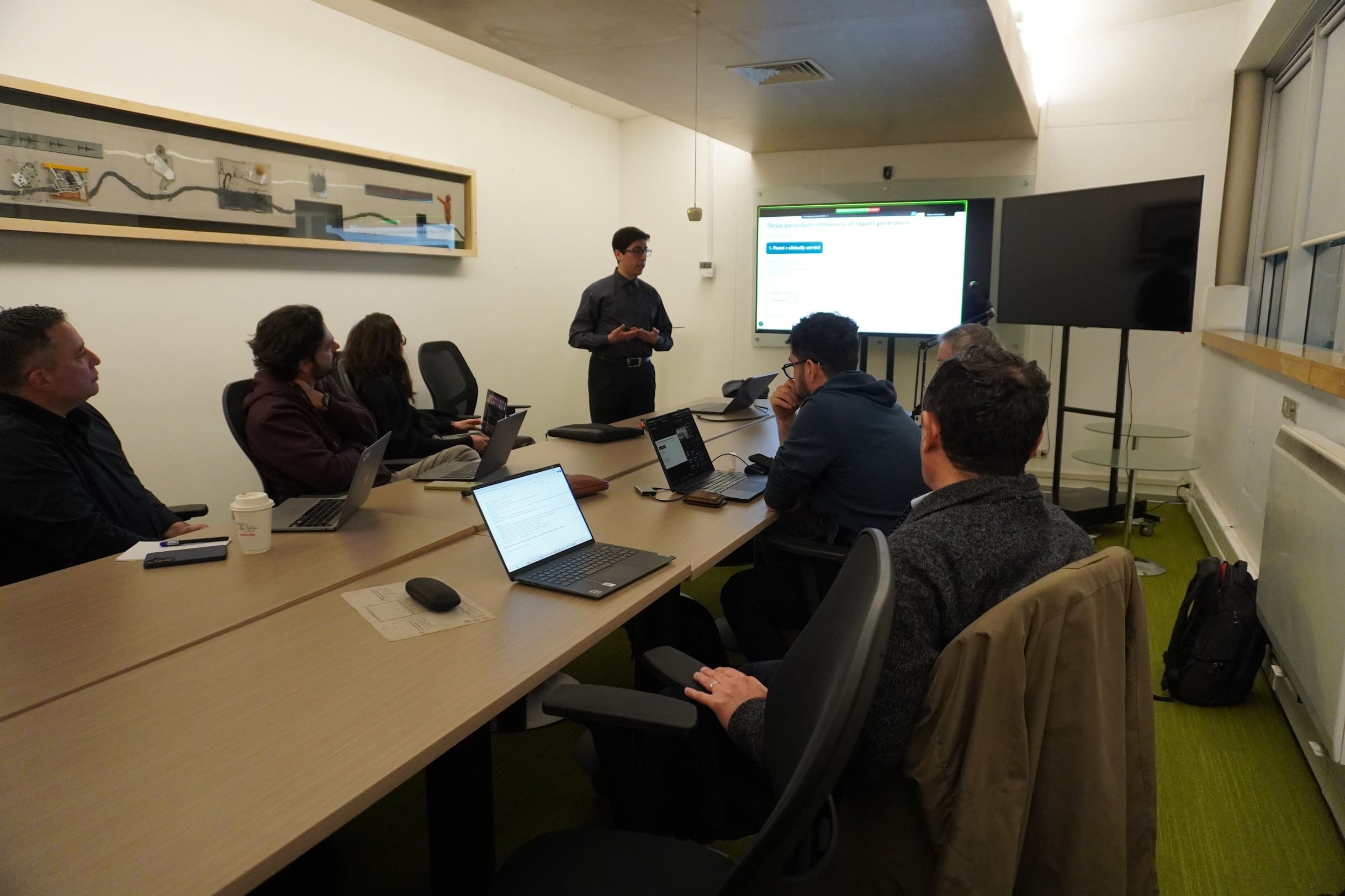
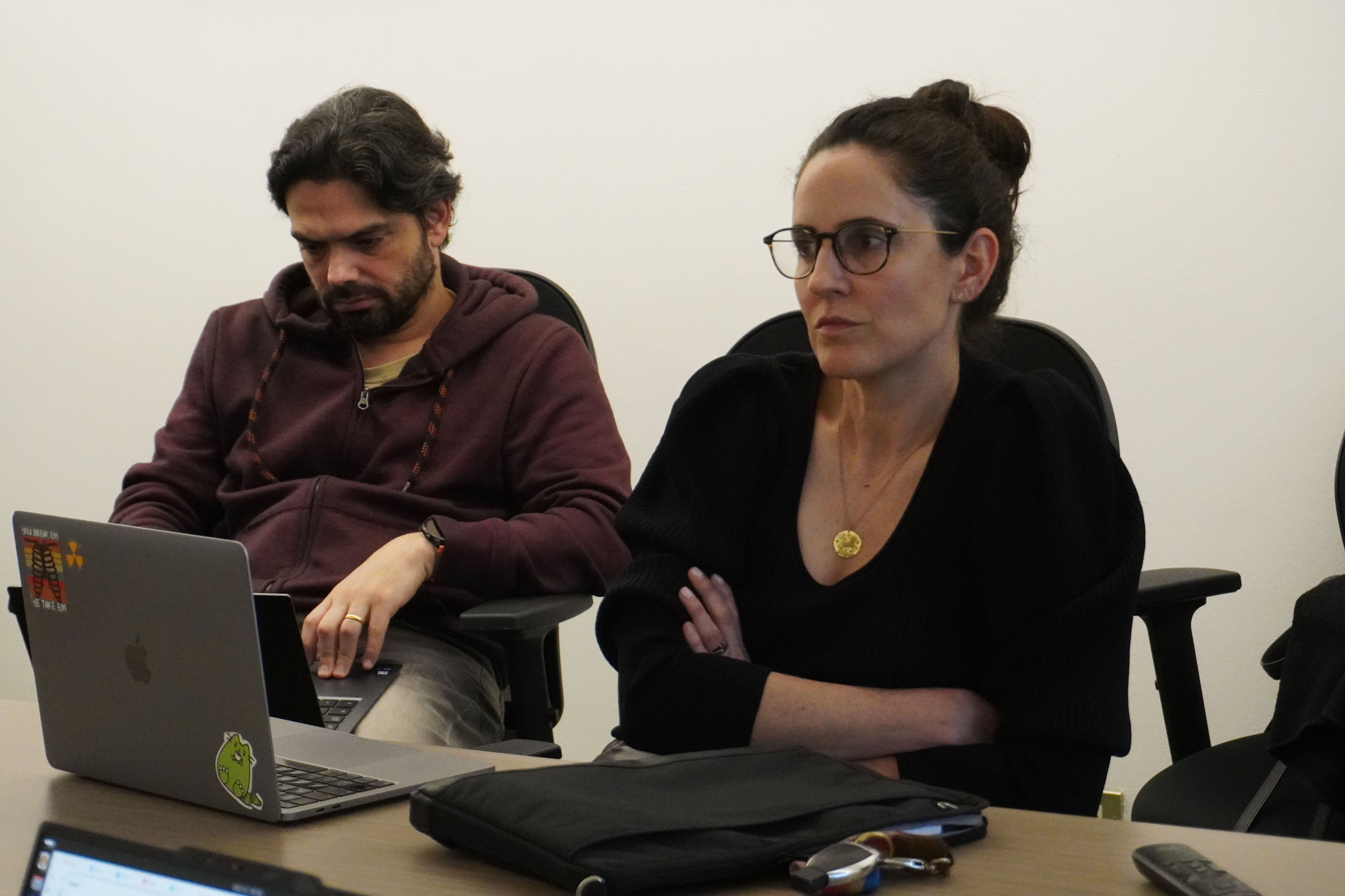
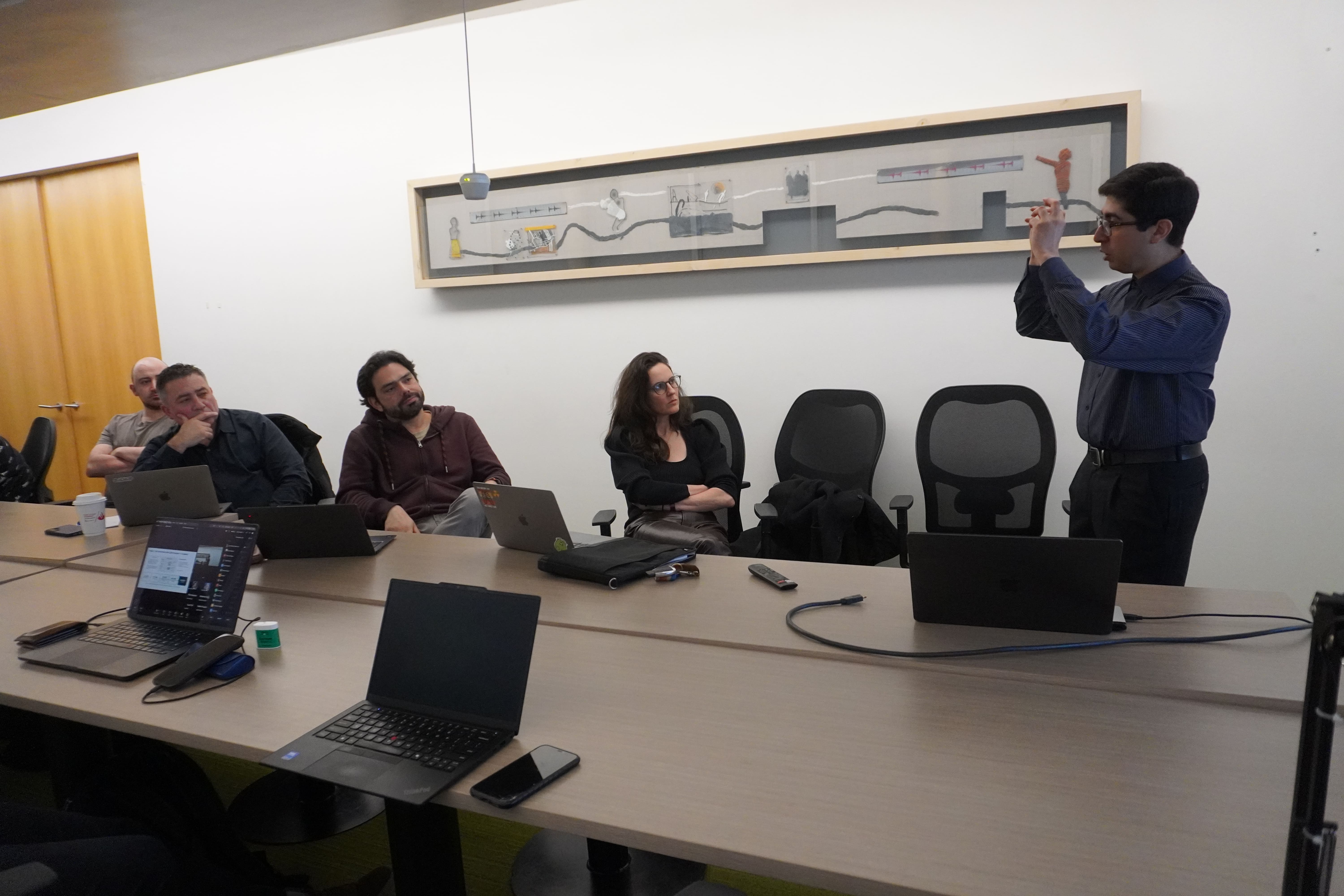
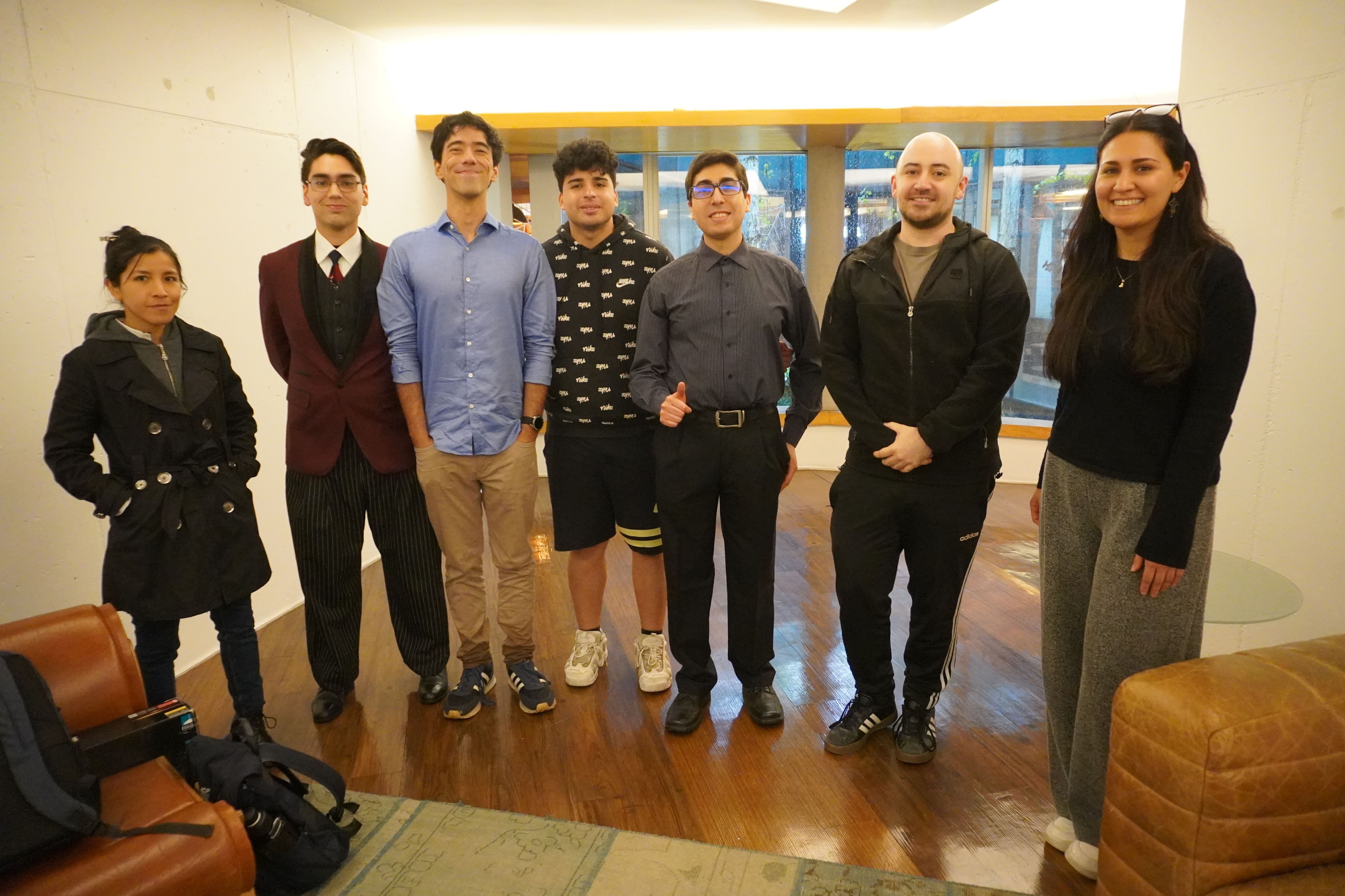
This approach guided six years of research and was the core of the defense that Pablo Messina Gallardo presented and passed on Tuesday, October 13, at the UC San Joaquín Campus. The thesis, titled “Automatic Generation of Radiological Reports Using a Visual Question Answering Approach,” was supervised by professors Álvaro Soto and Denis Parra, the latter an iHEALTH researcher. The committee comprised Hans Löbel and radiologist Cecilia Besa (UC), Rodrigo Salas (Universidad de Valparaíso), and René Vidal (University of Pennsylvania), and was chaired by Sergio Gutiérrez (UC).
For Parra, the value of the approach is clear: “The system generates the report, but it can also tell you which part of the image it looked at to do so. That provides more safety, more reliability, and more interpretability.”
The thesis is organized into three chapters, each based on a publication. The first, a systematic review of 40 studies published in ACM Computing Surveys (2022), showed that most systems were evaluated only with metrics that count words shared between two texts, without measuring whether the report was clinically correct. The survey was co-authored by iHEALTH researchers Claudia Prieto, Marcelo Andía, Sergio Uribe, Cristian Tejos, and Cecilia Besa.
“There were no good metrics to know whether an AI-generated report was good or not,” Parra explains. That is why the second chapter, presented at Findings of ACL 2024, proposes representing reports as clinical facts that are extracted and compared one by one. This gave rise to CXRFEScore, an open metric that can tell when two sentences say the same thing and when they contradict each other. The third chapter takes the step to the image with CURE, a training method that anchors each answer to a region of the X-ray and was selected for an oral presentation at CVPR 2026. Of the more than 16,000 papers submitted this year, only 141 received that distinction, less than 1%.
Teaching AI what healthy looks like
CURE starts from a diagnosis: the datasets used to train these models only mark image regions that show some abnormality. “So the model unintentionally learns that ‘pointing at something’ means ‘finding something wrong,’” Messina explains. For example, a reference model from Microsoft described healthy clavicles as fractured.
The solution combined two strategies: training the model to locate and describe anatomical regions, whether healthy or diseased, and applying a training plan that periodically identifies where it makes the most mistakes and has it practice on those. As a result, the average rate of invented findings dropped from 26.5% to 8.8%. For clavicles, it fell from around 60% to 1%.
True to an open-science approach, Messina released CXRFEScore as a tool that can be installed with a single command (pypi.org/project/cxrfescore). “I think it’s good practice for research not to stay in the paper, but to end up as a product that anyone can download and try in minutes,” he says.
He also highlights the interdisciplinary work: “This is a problem that can’t be solved from computer science alone. At iHEALTH, I was fortunate to work with radiologist Cecilia Besa, who helped me understand what a radiologist really needs from a report.” The thesis also benefited from the support of CENIA and from international collaborations. These include a research stay with René Vidal at Johns Hopkins, which continued at the University of Pennsylvania, and a remote internship in Bernard Ghanem’s lab at KAUST (Saudi Arabia), where CURE was born.
“Through his work at CENIA and iHEALTH, Pablo was able to show that it’s possible to have a very high impact worldwide,” Parra says.
Messina will now begin a postdoc at the University of Pennsylvania with René Vidal, who holds a joint appointment in Radiology and Electrical and Systems Engineering. There, he will continue working on trustworthy artificial intelligence for medical imaging and explore new challenges that combine brain data with video.