nnU-Net learning model achieves greater efficiency in myocardial segmentation
A study compared two advanced deep learning models — nnU-Net and MA-SAM — for automatic myocardial segmentation. While both achieved comparable segmentation quality, nnU-Net demonstrated notably superior computational efficiency: shorter training time and inference 490 times faster than manual segmentation.
Myocardial tissue characterization through T1 and T2 mapping by magnetic resonance imaging is a key tool for the non-invasive assessment of cardiovascular diseases. Myocardial segmentation — that is, the precise delineation of cardiac structures in images — is the essential first step for extracting quantitative tissue information.
However, these studies generate large datasets that require considerable time and clinical expertise to analyze. Faced with this challenge, artificial intelligence, and deep learning in particular, emerges as a promising solution to automate and accelerate this process.
A new article published in the journal NMR in Biomedicine evaluated two neural network architectures: nnU-Net, based on convolutional networks (CNN), and MA-SAM, based on Transformers. Both models were trained and evaluated using a dataset of 55 subjects, including healthy volunteers and patients with suspected cardiovascular disease.
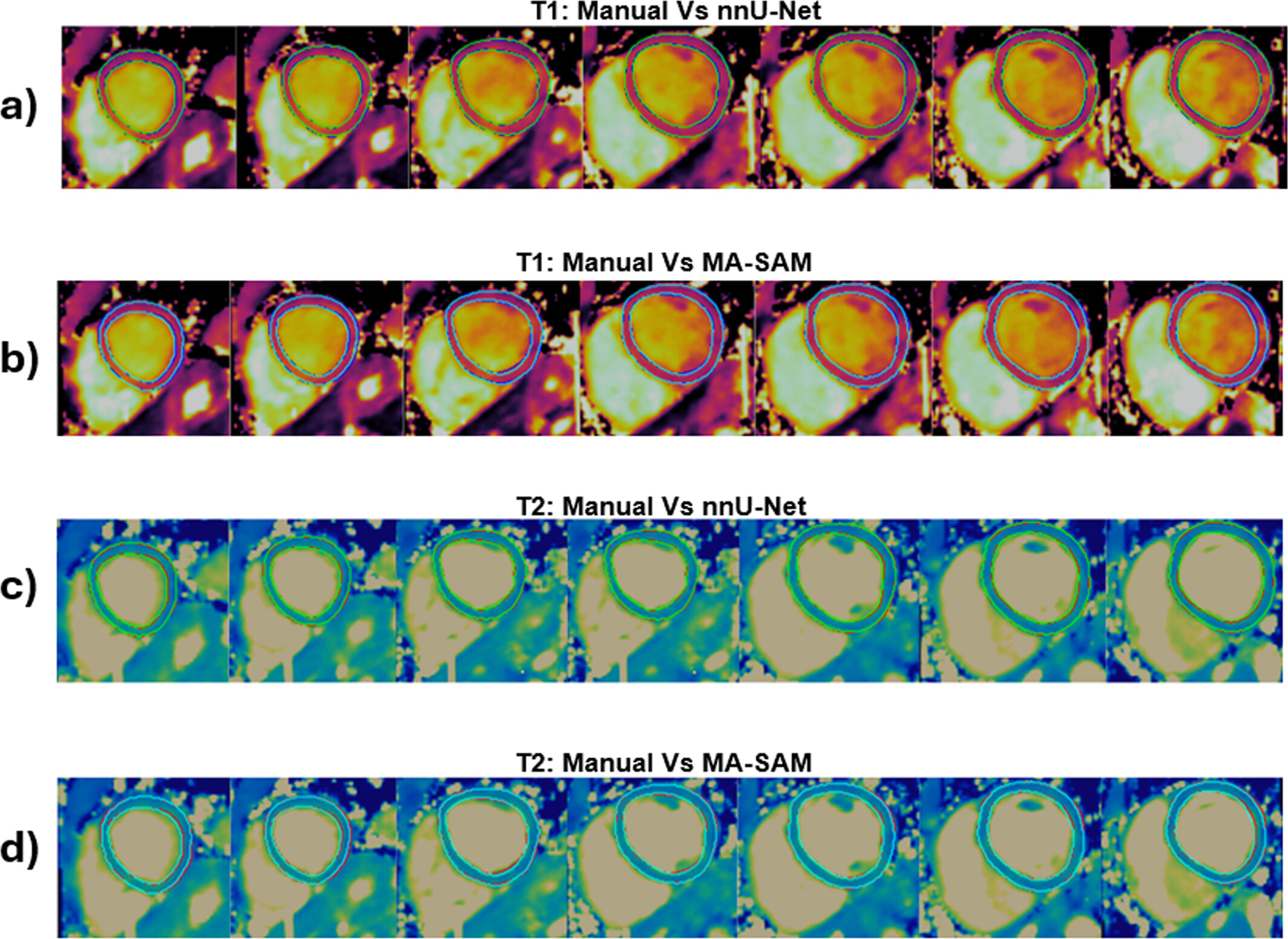
Results showed that both models produce visually similar, high-quality segmentations compared to the manual reference, though with notable differences in computational efficiency. nnU-Net completed training in 5 hours versus the 9 hours required by MA-SAM, and segmented a full 3D map in just 3.7 seconds, compared to 76 seconds for its counterpart. Compared to manual segmentation — which takes approximately half an hour — nnU-Net was 490 times faster and MA-SAM 23 times faster.
"The work sought to compare frameworks already existing in the literature, and it was concluded that both convolutional networks and transformers can achieve similar results in terms of segmentation quality. However, transformer-based models continue to require considerably higher computational cost," said Carlota Rivera, a doctoral student at iHEALTH and one of the study's authors.
Clinical implications and next steps
The researcher also highlighted the clinical potential of these findings: "The faster the image analysis, the better the clinical workflow can be. Automating this process could accelerate the extraction of relevant parameters and potentially contribute to faster, more efficient diagnosis."
For this technology to be implemented in hospitals or medical centers, it will be necessary to validate the models in larger and more diverse cohorts, and to ensure that results are robust across different clinical contexts.
Among future research directions, the team is considering evaluating the performance of these models on 0.55 T MRI scanners — this study was conducted at 1.5 T — and exploring semi-supervised learning methods, which would allow robust models to be trained with fewer manual segmentations, reducing the time and effort required to generate training data.
Rivera GC, Hua A, Botnar RM, Prieto C. Deep Learning Myocardial Segmentation in 3D Whole-Heart Joint T1/T2 Mapping: Comparison of nnU-Net and MA-SAM. NMR Biomed. 2026 Apr;39(4):e70252. doi: 10.1002/nbm.70252. PMID: 41790043.