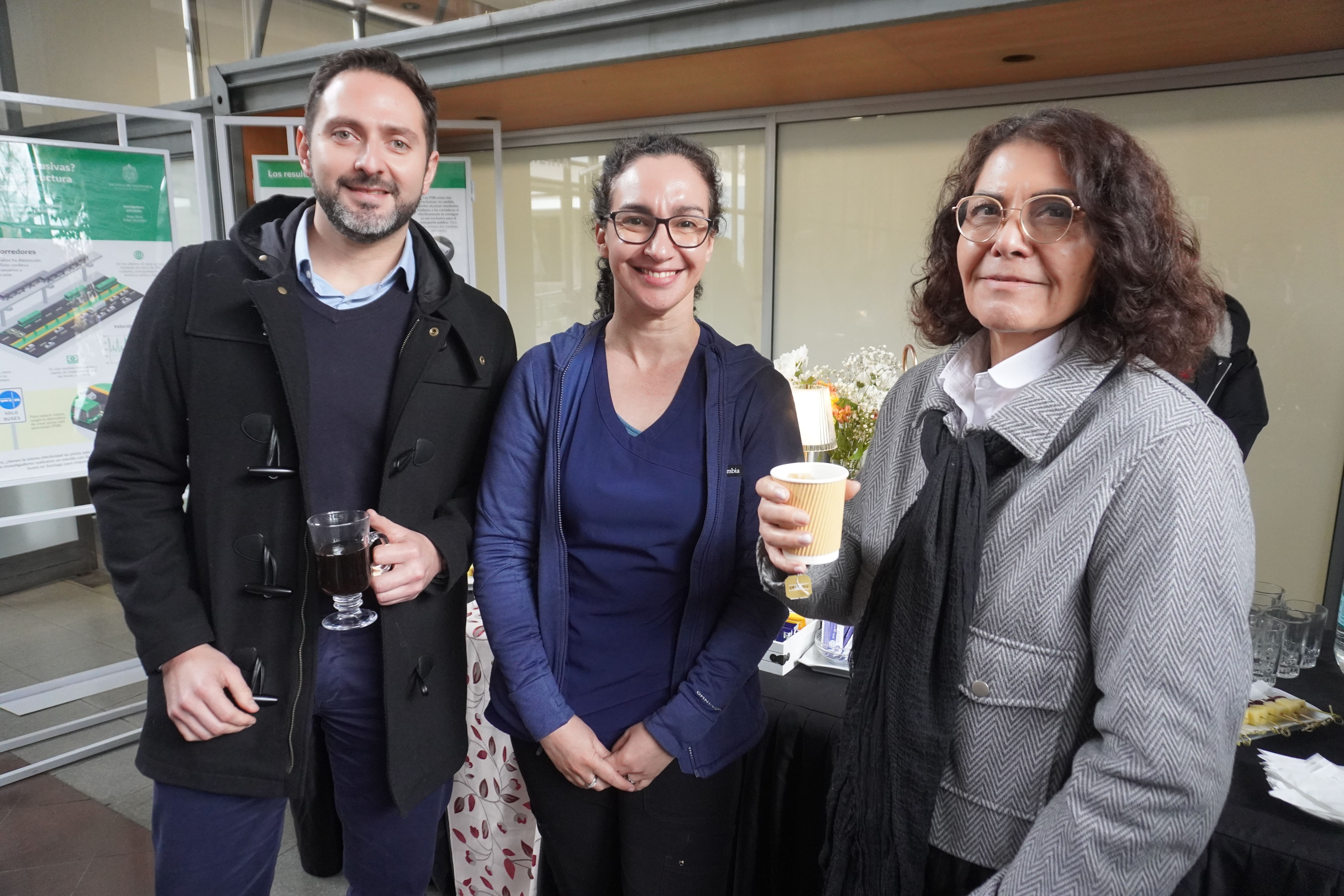
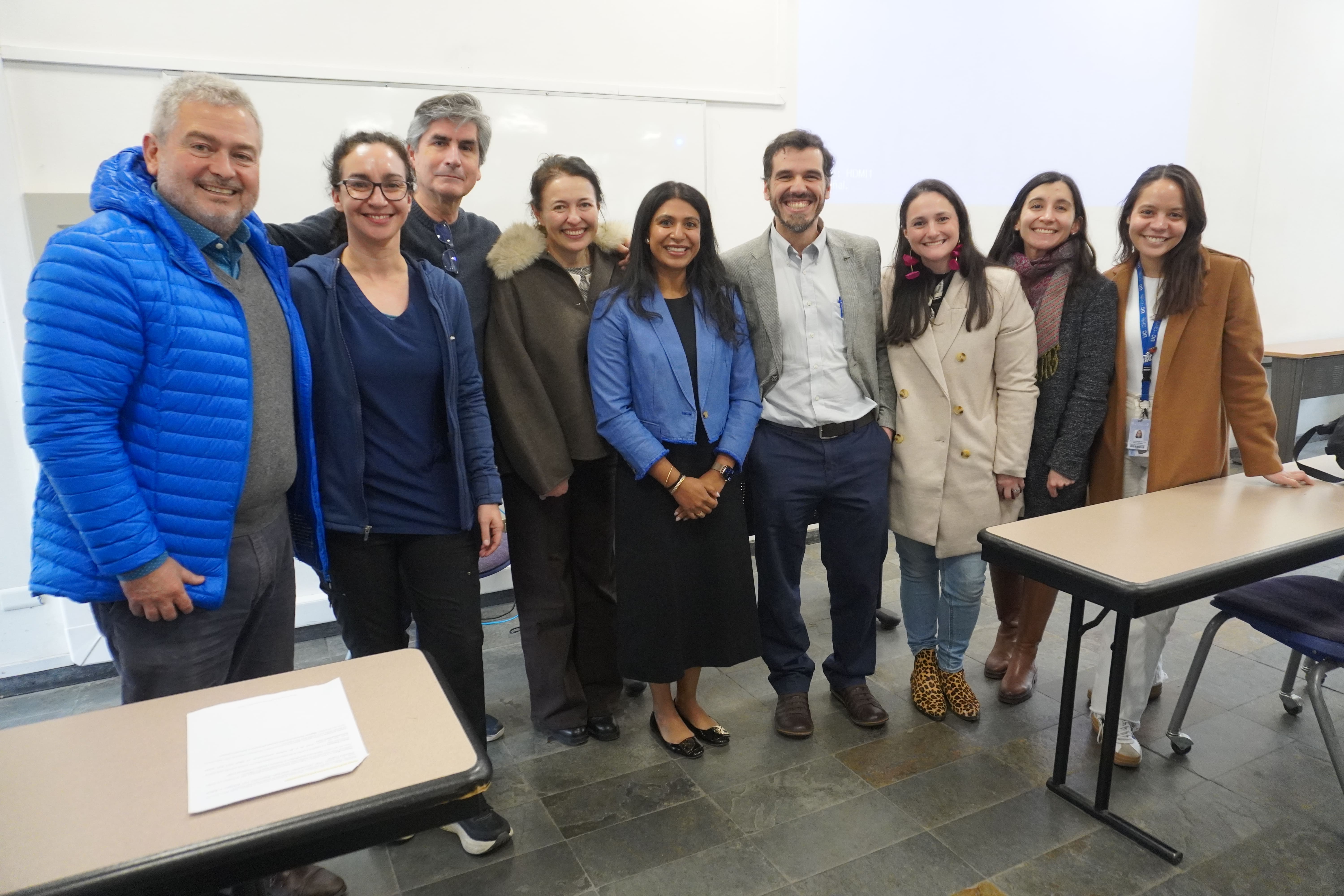
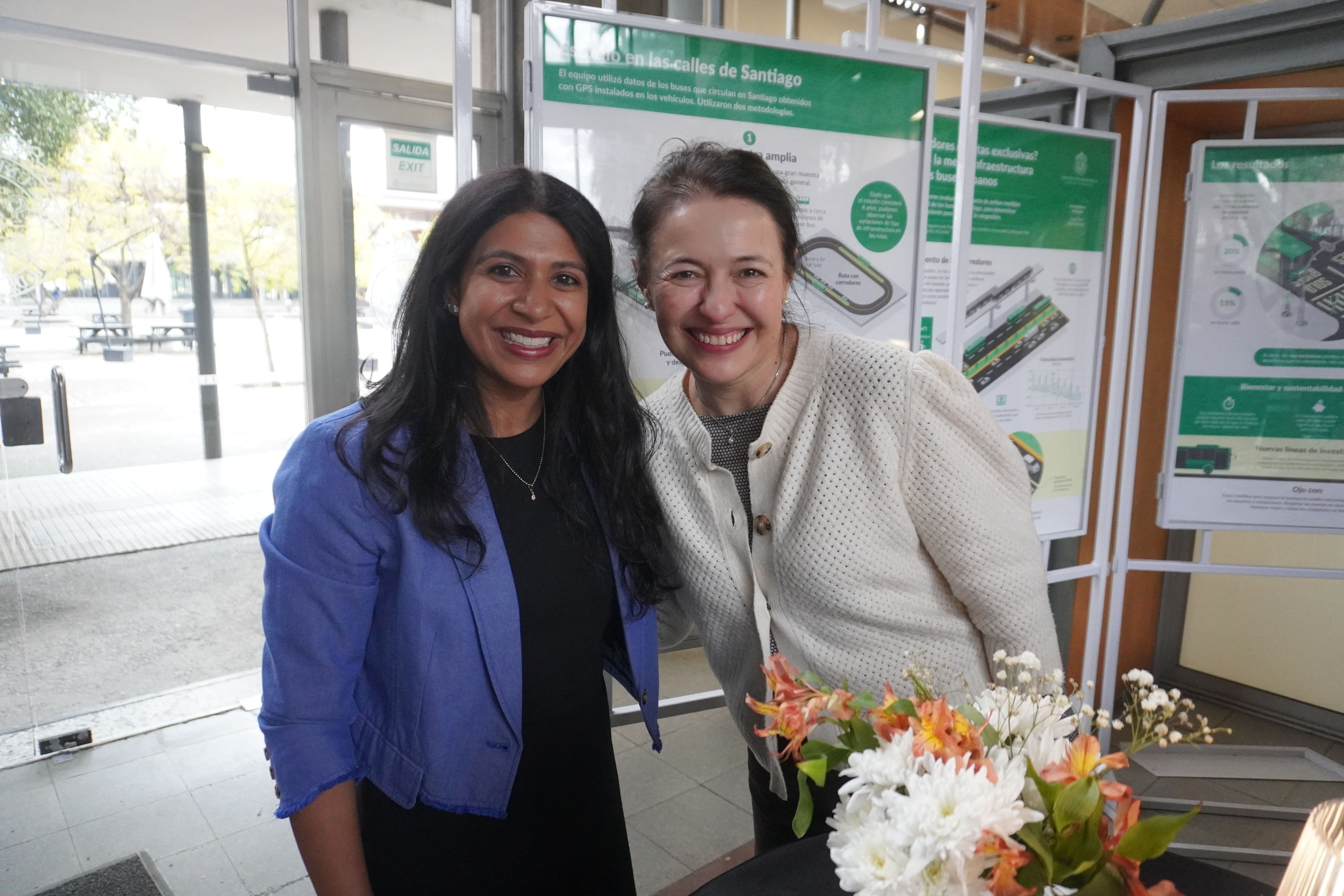
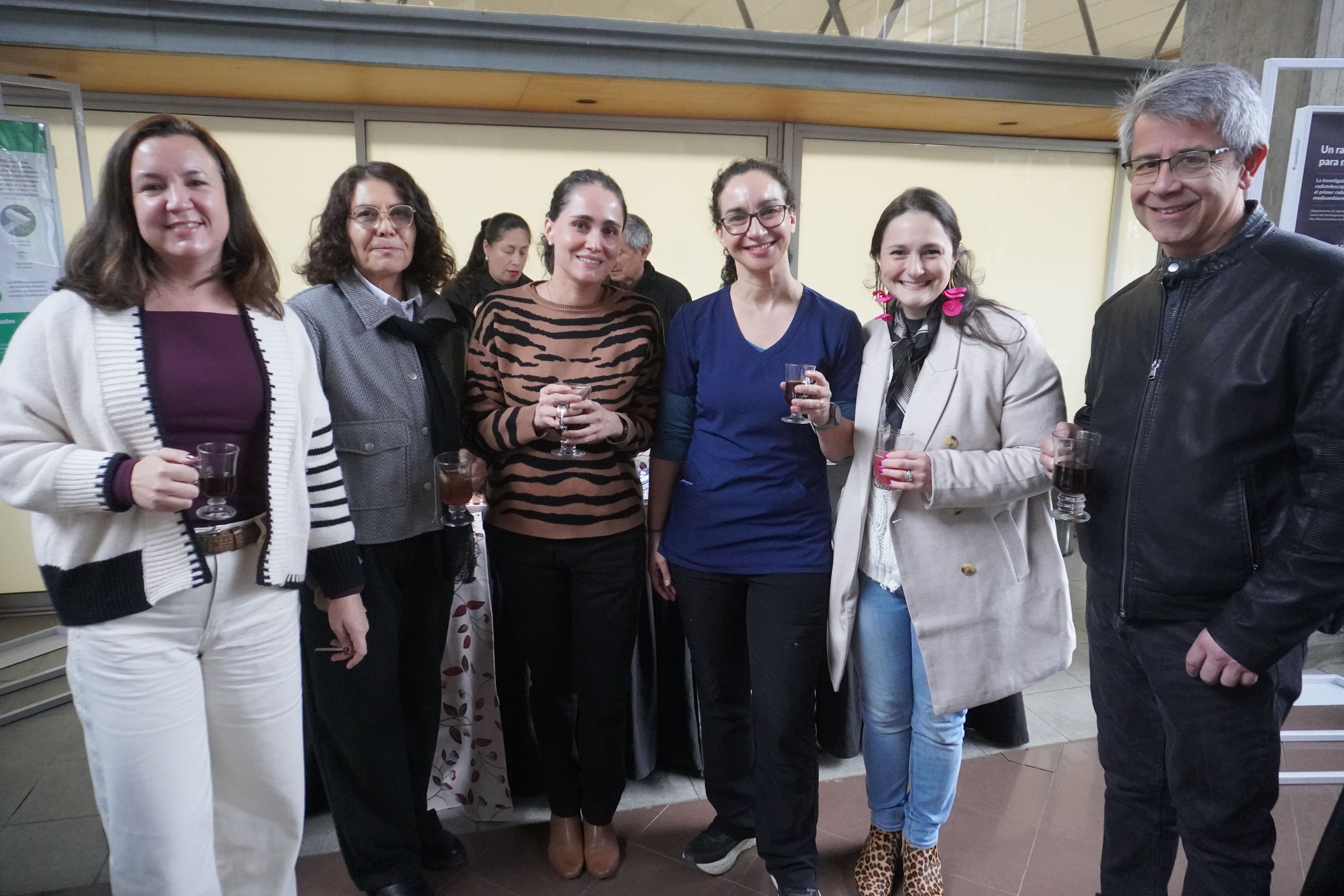
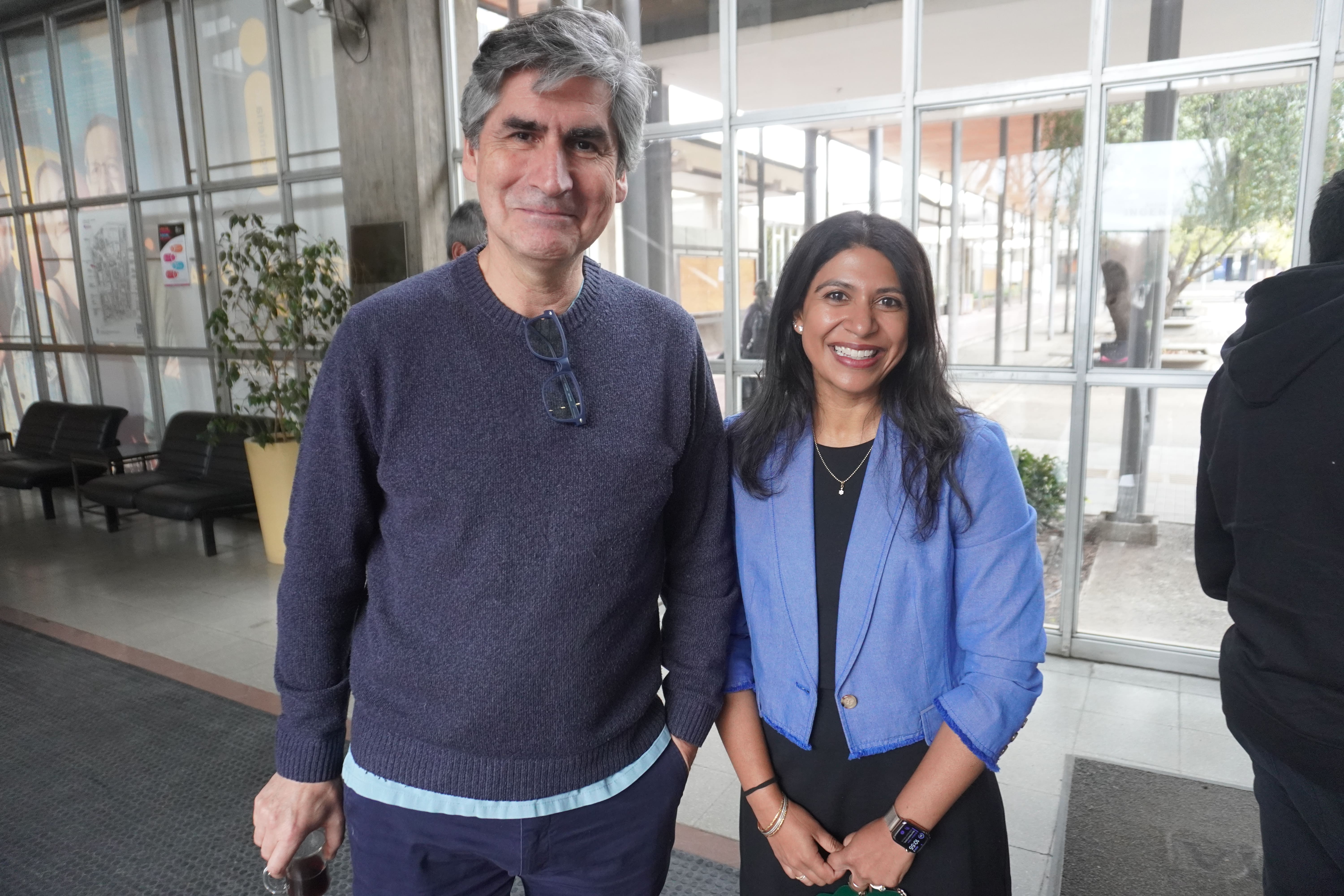
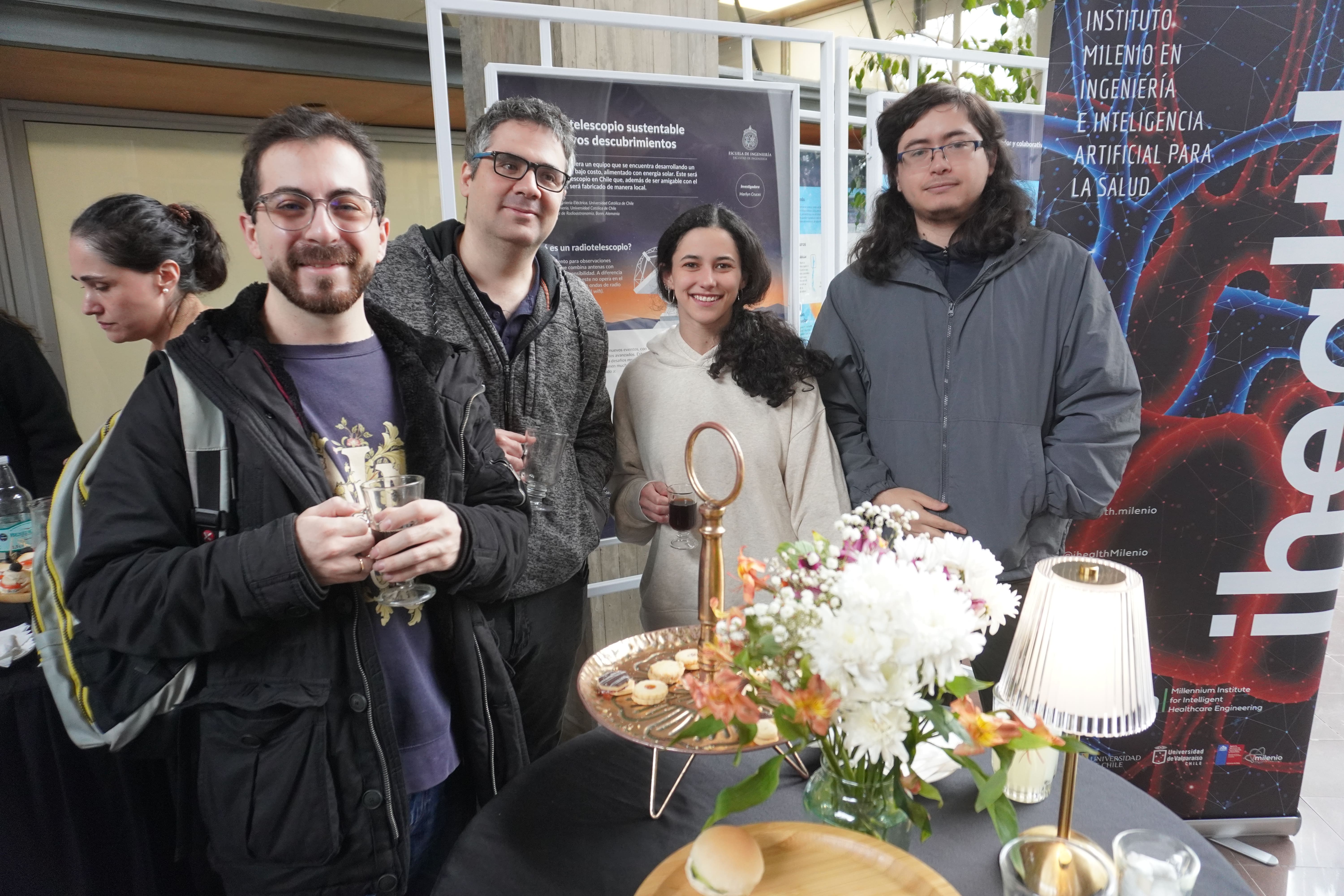
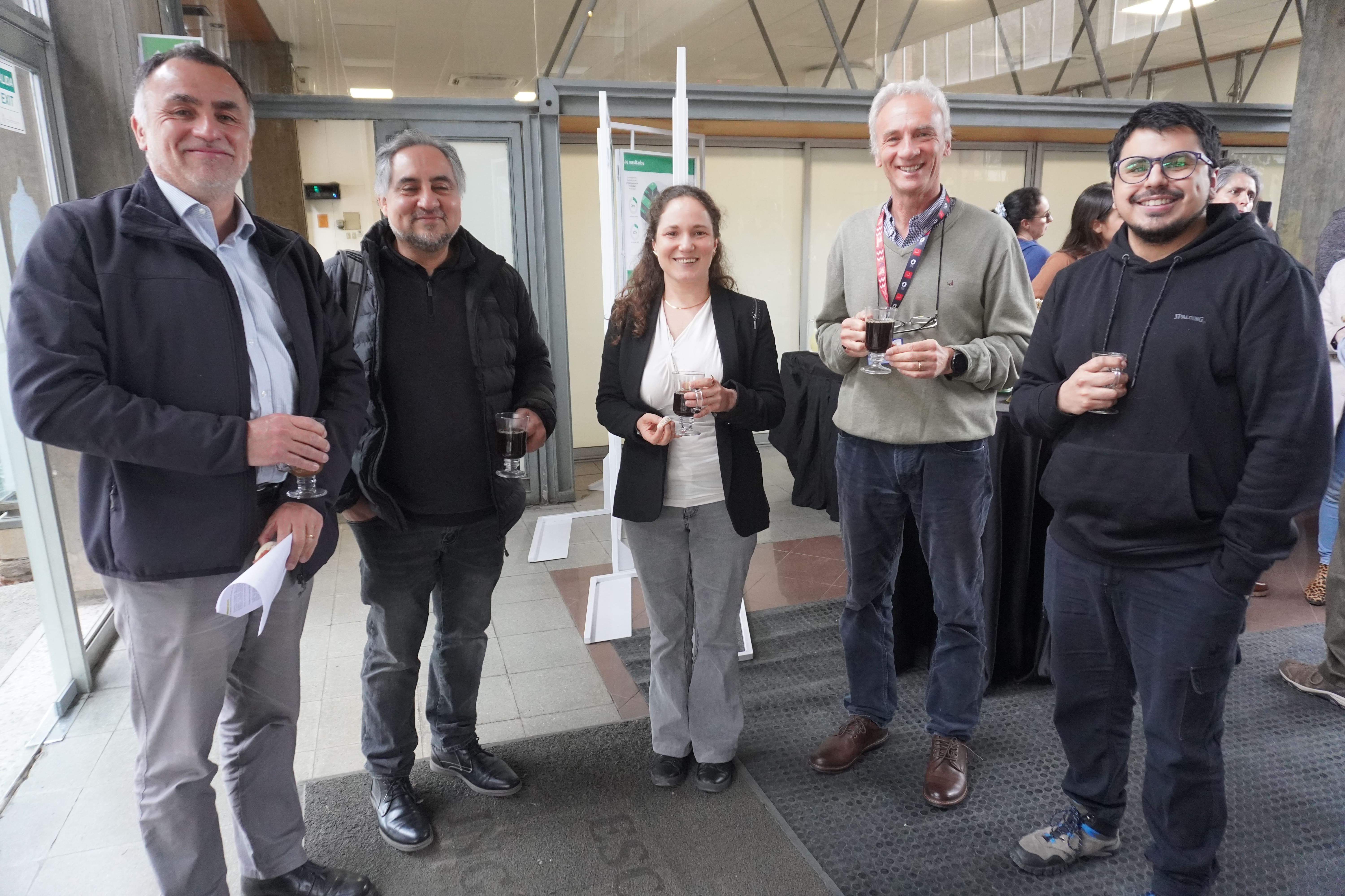
Medical imaging experts gathered at the iHEALTH Ultrasound Imaging Symposium
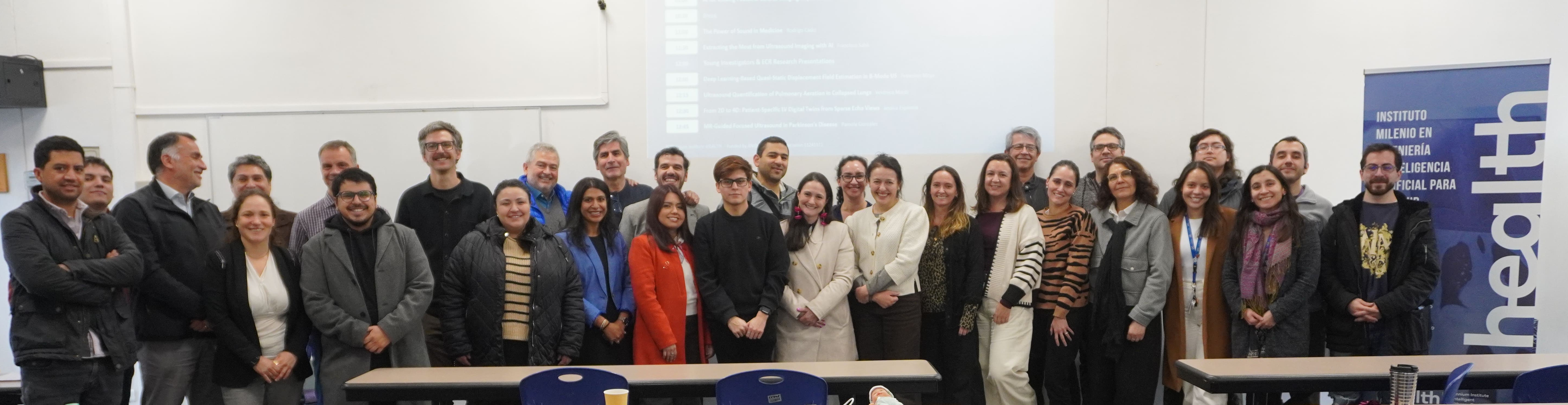
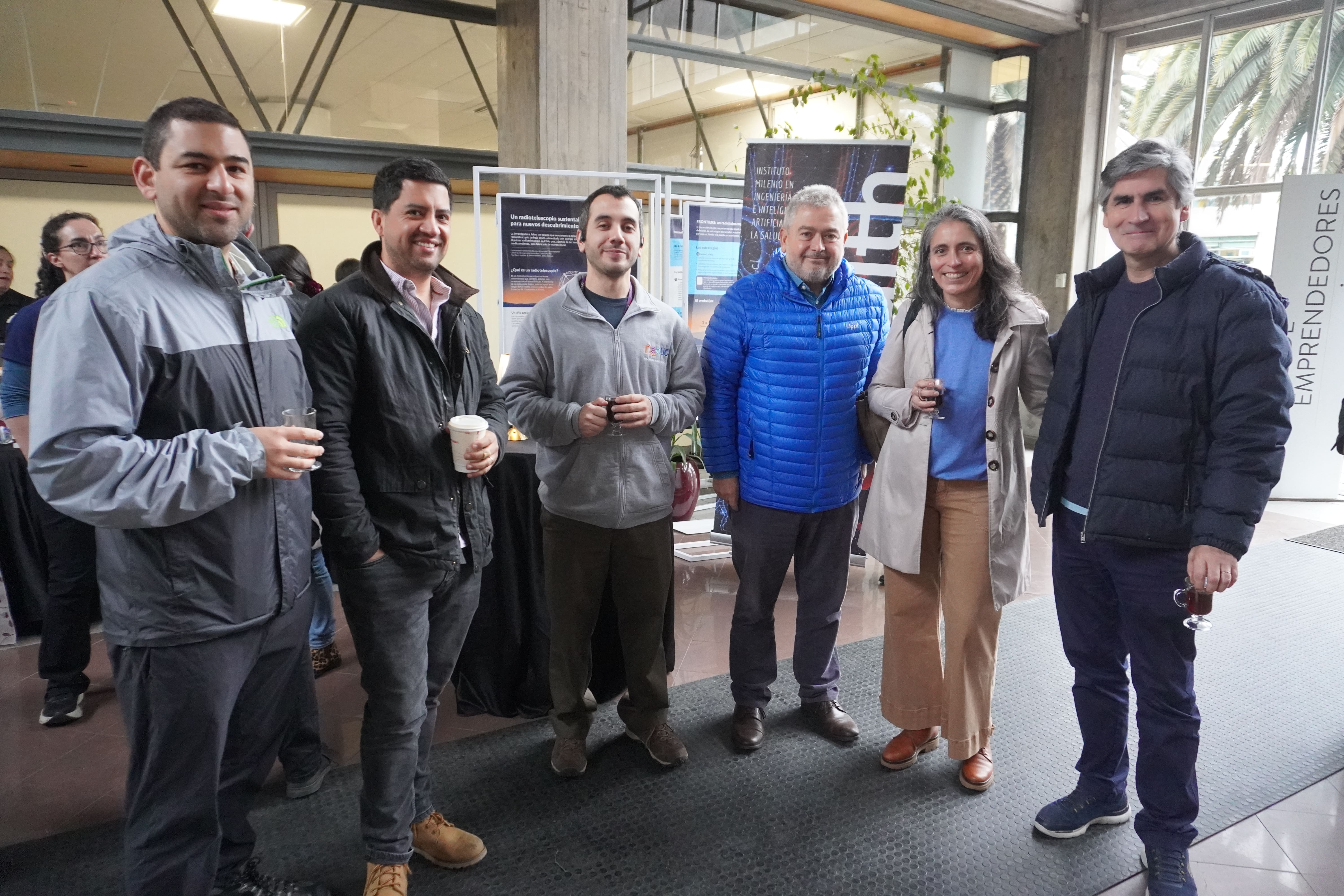
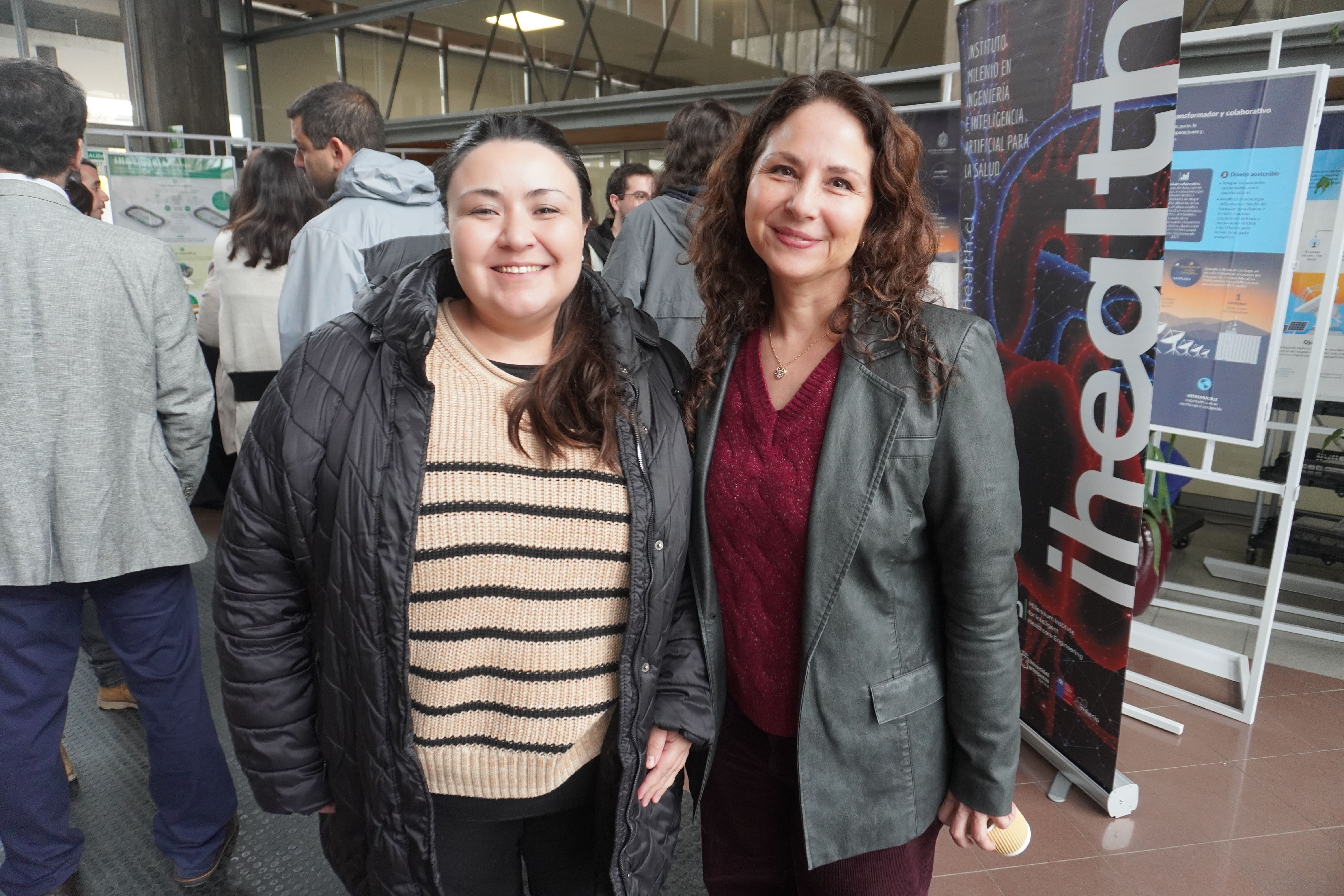
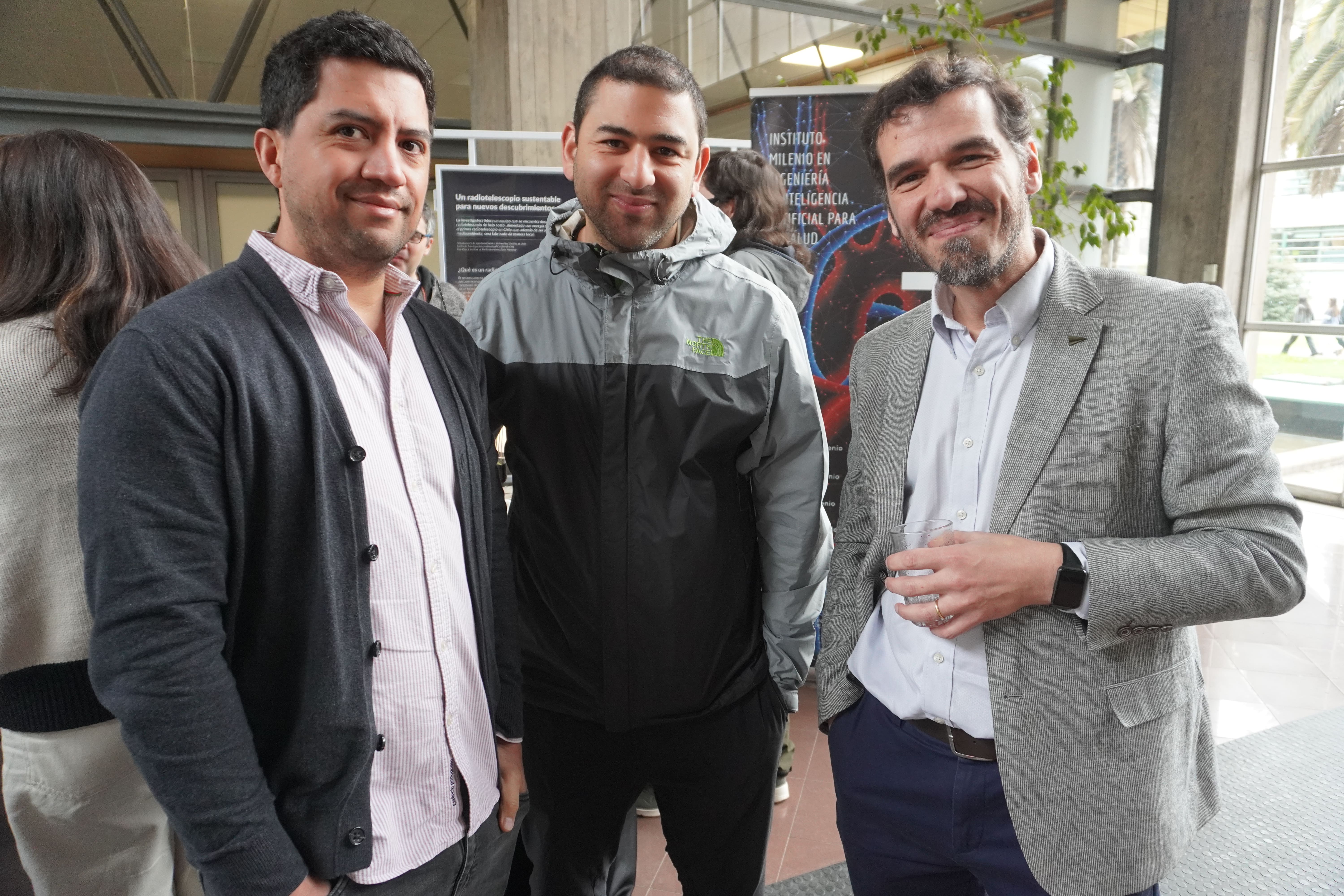
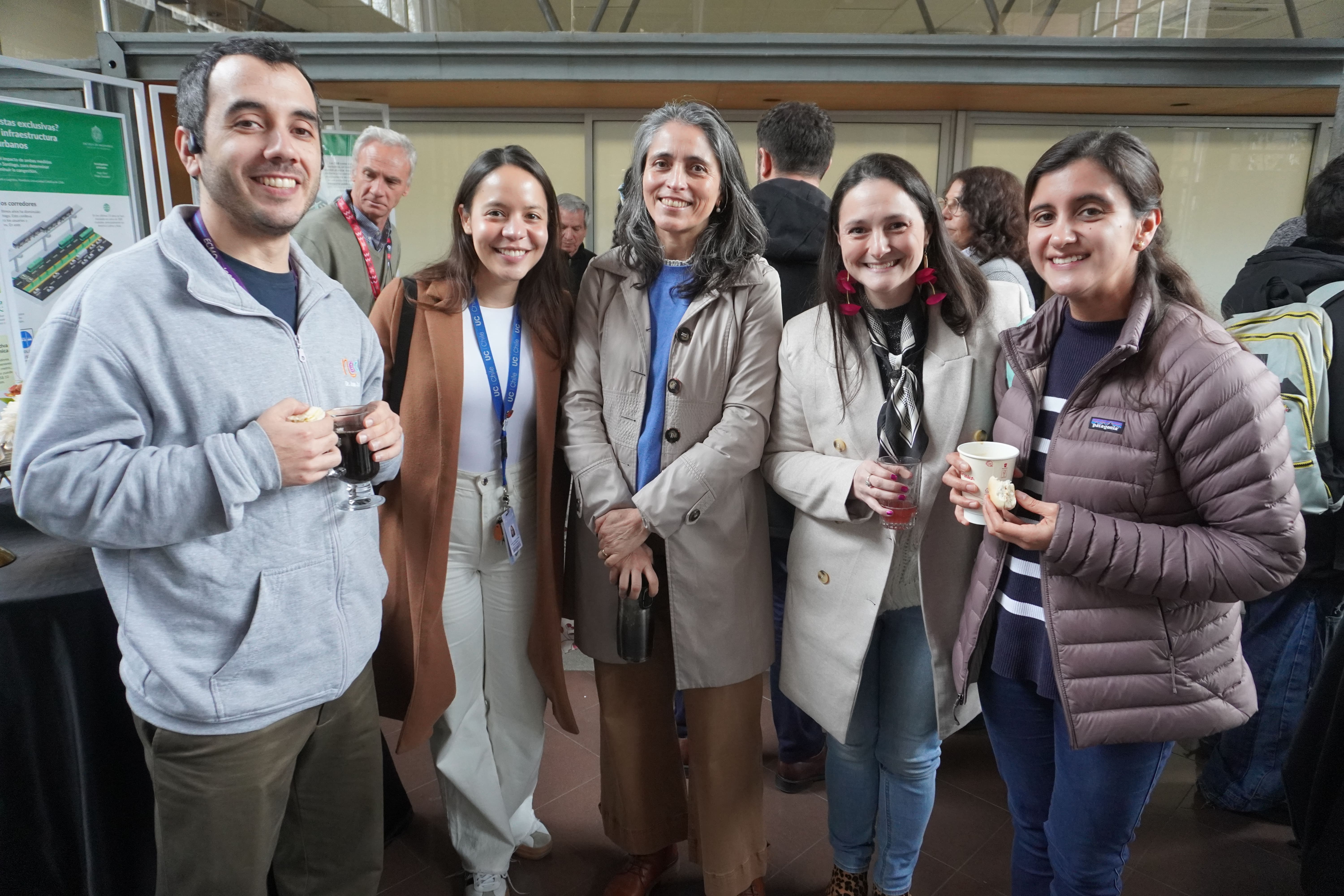
On Monday, August 3, the Raúl Devés Study Hall at UC's San Joaquín Campus was the meeting point for the iHEALTH Ultrasound Imaging Symposium: Advances and Future Outlook, a day organized by the Millennium Institute iHEALTH that brought together researchers, clinicians, and students around a central idea: how to extract more information from one of the most accessible and versatile imaging technologies in medicine.
As one of the day's talks noted, nearly two-thirds of the world's population lacks access to diagnostic imaging, even though 70 to 80% of clinical questions can be answered with basic X-ray or ultrasound. Because it uses no ionizing radiation, is portable, works in real time, and costs a fraction of a CT or MRI, ultrasound is the modality with the greatest potential to close that gap. The program was built on this premise, spanning pediatric cardiology, artificial intelligence, signal physics, neurology, and iHEALTH's own research lines.
The welcome was given by Marcelo Andia, Alternate Director of iHEALTH, who introduced the day and its interdisciplinary spirit before giving way to the opening keynote, "From Pixels to Predictions: AI in Pediatric Echocardiography," delivered by Dr. Charitha Doodipala Reddy, Clinical Associate Professor in the Division of Pediatric Cardiology at Stanford University and Lucile Packard Children's Hospital.
Drawing on a real clinical case, Reddy posed the question that ran through much of the day: within a few years, algorithms could interpret pediatric echocardiograms better than many experts, and the challenge lies in how clinicians learn, implement, and take responsibility for these tools. Her tour through AI-assisted acquisition, view classification, automated ventricular function measurement, and risk stratification closed with an idea that resonated in the room: "algorithms see pixels, doctors see patients; algorithms must be brilliant, and clinicians wise."
Dr. Guillermo Larios, pediatric cardiologist and Associate Professor at the Pontificia Universidad Católica de Chile, brought that discussion home to the national context with "AI for Closing Pediatric Cardiac Imaging Gaps in Chile." Larios laid out the concrete gaps in Chilean pediatric cardiology—fetal diagnosis, imaging far from Santiago, and access to cardiac MRI—and defended a clear thesis: every solution must be validated locally and built on the infrastructure hospitals already have, such as electrocardiography and echocardiography. His closing invitation pointed to digital twins that combine echo and ECG, a field where clinicians and engineers can work in the same room.
Dr. Rodrigo Cádiz, Full Professor in the Department of Electrical Engineering and the Institute of Music at UC and adjunct researcher at iHEALTH, offered a different perspective in "The Power of Sound in Medicine." Starting from Laennec's invention of the stethoscope in 1816, Cádiz recalled that the body is a source of sound—valves, flow, airways, vocal cords—and that auscultation has remained stuck where Laennec left it: it is not continuous, portable, comparable, or quantitative. His talk explored how signal processing, sonification, and artificial intelligence can restore listening to its diagnostic value.
Closing the plenary block, Dr. Francisco Sahli, Assistant Professor in the Department of Mechanical and Metallurgical Engineering and the Institute for Biological and Medical Engineering at UC and principal investigator at iHEALTH, presented "Extracting the most from ultrasound imaging with AI." Sahli framed inverse problems in ultrasound within the argument of access: improving CT or MRI benefits the third of the world that already has them, while extracting more from a portable probe reaches the other two-thirds. That asymmetry, he argued, is what gives meaning to the entire effort.
Early Career Researchers
The day devoted a block to presentations by young researchers, beginning with Francisco Moya, a Master's student in Mechanical Engineering Sciences at Universidad Técnica Federico Santa María, supervised by Drs. Julio Sotelo and Joaquín Mura, who presented "Deep Learning-Based Quasi-Static Displacement Field Estimation in B-Mode Ultrasound Imaging." His work addresses elastography, a technique that characterizes the mechanical properties of tissues—key to distinguishing healthy from pathological tissue—through a deep learning model that estimates 2D displacement fields directly from B-mode images before and after compression, trained with synthetic data generated by simulation.
Verónica Marín, a doctoral student in Engineering Sciences (Electrical Engineering) at UC, presented "Ultrasound Quantification of Pulmonary Aeration in Collapsed Lungs." Marín introduced a methodology to quantify pulmonary aeration from ultrasound signals using parameters such as total intensity and Shannon entropy, with the aim of objectively guiding alveolar recruitment maneuvers. To this end, she developed a collapsed-lung phantom that reproduces different degrees of aeration in a controlled way.
Jessica Espinoza, Research Engineer at iHEALTH under the supervision of Dr. Francisco Sahli, presented "From 2D to 4D: building patient-specific LV digital twins from sparse echocardiographic views." Her work transforms routinely acquired 2D echocardiographic views into a dynamic, three-dimensional model of the left ventricle specific to each patient, with particular value in the pediatric population. The pipeline combines automatic segmentation of the endocardial border, estimation of each view's position in space, and reconstruction of the cardiac shape throughout the cycle, achieving promising results in pediatric patients.
The block closed with Pamela González, a doctoral student in Medical Sciences at UC, who presented "Magnetic Resonance Guided Focused Ultrasound in Parkinson's Disease." González presented the MRgFUS technique, which combines high-intensity focused ultrasound with magnetic resonance to perform brain procedures without open surgery, guiding treatment with anatomical imaging and real-time MR thermometry. In Parkinson's disease, this minimally invasive approach opens new therapeutic possibilities for patients who continue to experience disabling symptoms despite available treatments.
A day that reflects iHEALTH's vision
The symposium captured the purpose that drives the Millennium Institute iHEALTH: to develop solutions that integrate engineering and artificial intelligence to improve medical image-based healthcare, making it in the long term more precise, efficient, effective, and accessible for all Chileans. From pediatric cardiac diagnosis to neurology, and from signal physics to digital twins, the day showed how interdisciplinary work between clinicians and engineers makes it possible to extract the full potential of a technology as widespread as ultrasound.