iHEALTH releases a cardiac imaging database acquired with low-field magnetic resonance imaging to the scientific community
The dataset includes scans from 110 healthy volunteers performed on a 0.55 T clinical scanner, and is available in open access through the Zenodo platform.
The Millennium Institute iHEALTH has published a new cardiac imaging database on Zenodo, an open-access repository developed under the European OpenAIRE program and operated by the European Organization for Nuclear Research (CERN), which enables researchers around the world to share data for research purposes. This initiative builds on the knee imaging database the Institute made available to the scientific community some time ago.
The dataset contains scans from 110 healthy volunteers, acquired on iHEALTH’s 0.55 T low-field MRI scanner. This makes it a larger-scale resource, aimed at increasing data availability in the field of cardiovascular medical imaging.
For Claudia Prieto, Director and Principal Investigator of iHEALTH, this new dataset is concrete evidence of the Institute’s progress in this line of work: “When we published the knee database, we said it was a starting point, and today we are taking an important step forward. Having cardiac images from 110 volunteers acquired on a low-field system, which is more accessible than conventional scanners, opens up new opportunities to develop and validate reconstruction, analysis and artificial intelligence methods applied to cardiovascular health.”
She added: “Unlike most available databases, which only provide already reconstructed images, this dataset also includes the raw acquisition data, allowing other groups to work from the origin of the signal and compare their own reconstruction methods.”
Cardiovascular diseases remain one of the leading causes of death in Chile and worldwide. “That is why we believe it is essential to have high-quality open data that allows researchers in different countries to advance diagnostic tools that may, in the future, reach more people, especially in places where access to magnetic resonance imaging is currently limited,” the Director noted.
The project was led by Claudia Prieto and René Botnar, iHEALTH researcher, as Project Leaders, while Ignacio Celis, medical technologist on the team, was responsible for building the database and performing the scans as Data Collector.
Data characteristics
Each of the 110 volunteers underwent the same scanning protocol on the low-field scanner, consisting of two types of acquisitions.
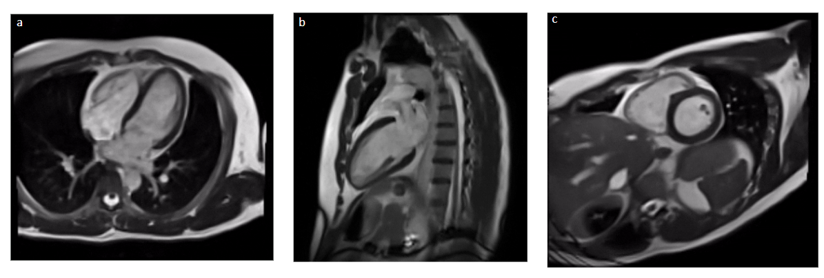
First, 2D cine images were obtained, which show the motion of the heart throughout the cardiac cycle, in three views: four-chamber, two-chamber and short-axis. These are the views routinely used in clinical practice to assess cardiac anatomy and function.
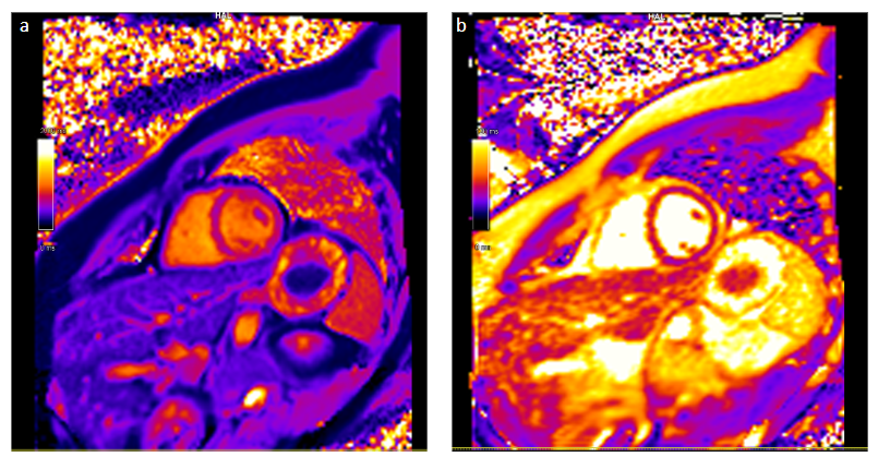
In addition, 2D T1 and T2 maps were acquired in the short-axis view. These quantitative techniques provide information about the tissue properties of the heart muscle and are useful for characterizing different pathologies.
It is worth noting that informed consent was obtained from all volunteers prior to the scans, along with ethics approval to share the images in anonymized form with the scientific community.
Link to the database: https://zenodo.org/records/23089164