iHEALTH Pilot Database Now on Zenodo
Data is already available online for researchers worldwide.
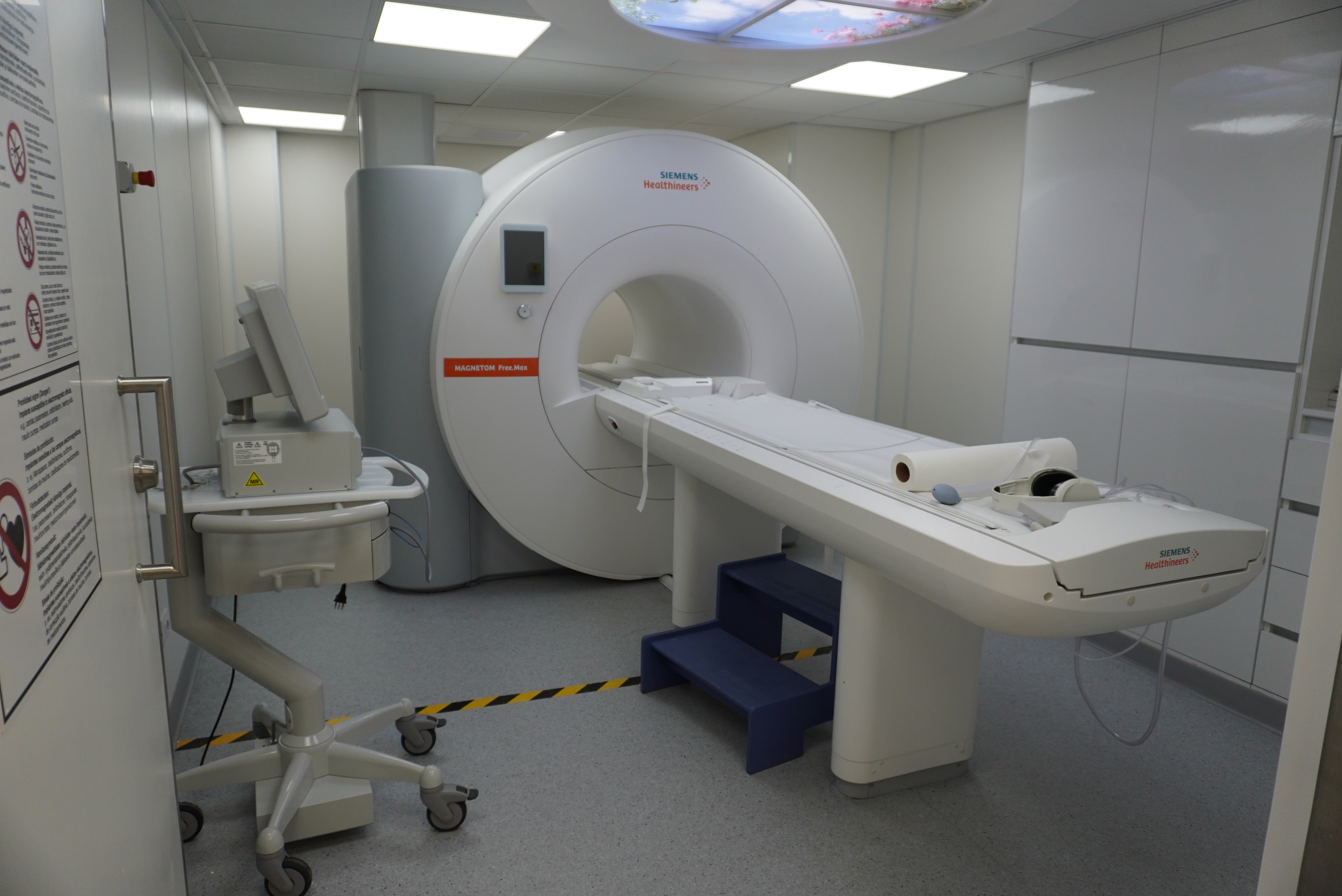
With the goal of contributing to data availability in the field of medical imaging, the Millennium Institute iHEALTH has made knee images available to the scientific community through Zenodo — an open-access repository developed under the European OpenAIRE program and operated by the European Organization for Nuclear Research (CERN). The images were acquired using a 0.55 T clinical MRI scanner and can now be used and shared by researchers around the world.
For Claudia Prieto, director and principal investigator of iHEALTH, publishing this type of information is a cornerstone of global scientific collaboration: "Making this information available to the scientific community is a significant step, as it allows us to share anonymized data acquired in Chile using an MRI scanner that is more accessible than conventional ones."
She added: "We are aware that this is a limited dataset of 20 subjects, so it should be understood as a starting point. However, it is a valuable resource for developing and validating new medical imaging and AI methods. We are currently working on acquiring larger-scale datasets, which will be essential for advancing this research further."
The work was carried out in collaboration with the team from the international project A4IM (Affordable, Accessible, Adaptable, and Accurate MRI), led by Christoph Kolbitsch from the German national metrology institute (PTB), of which iHEALTH's director is a member.
Dataset Characteristics
Ignacio Celis, a medical technologist at iHEALTH, scanned both knees of 20 recruited subjects using two types of sequences. First, a qualitative protocol was applied consisting of three consecutive 2D sequences with Cartesian trajectories and optimized parameters to obtain Proton Density (PD)-, T1-, and T2-weighted images, generating a total of 6 datasets.
The quantitative protocol was carried out with two image sets: a 2D Modified Look-Locker Inversion Recovery (MOLLI) (3-3-5) sequence to obtain a T1 map, and a T2-prepared balanced steady-state free precession (T2p-bSSFP) sequence to obtain a T2 map.
Images were acquired in the sagittal plane for better anatomical visualization of the knee, with a total scan time of just over 17 minutes per volunteer. Notably, informed consent was obtained from all volunteers prior to imaging, and ethical approval was granted to share the data in anonymized form with the scientific community.