Artificial intelligence to outline breast tumors in 3D: synthetic data as a starting point when clinical data are scarce
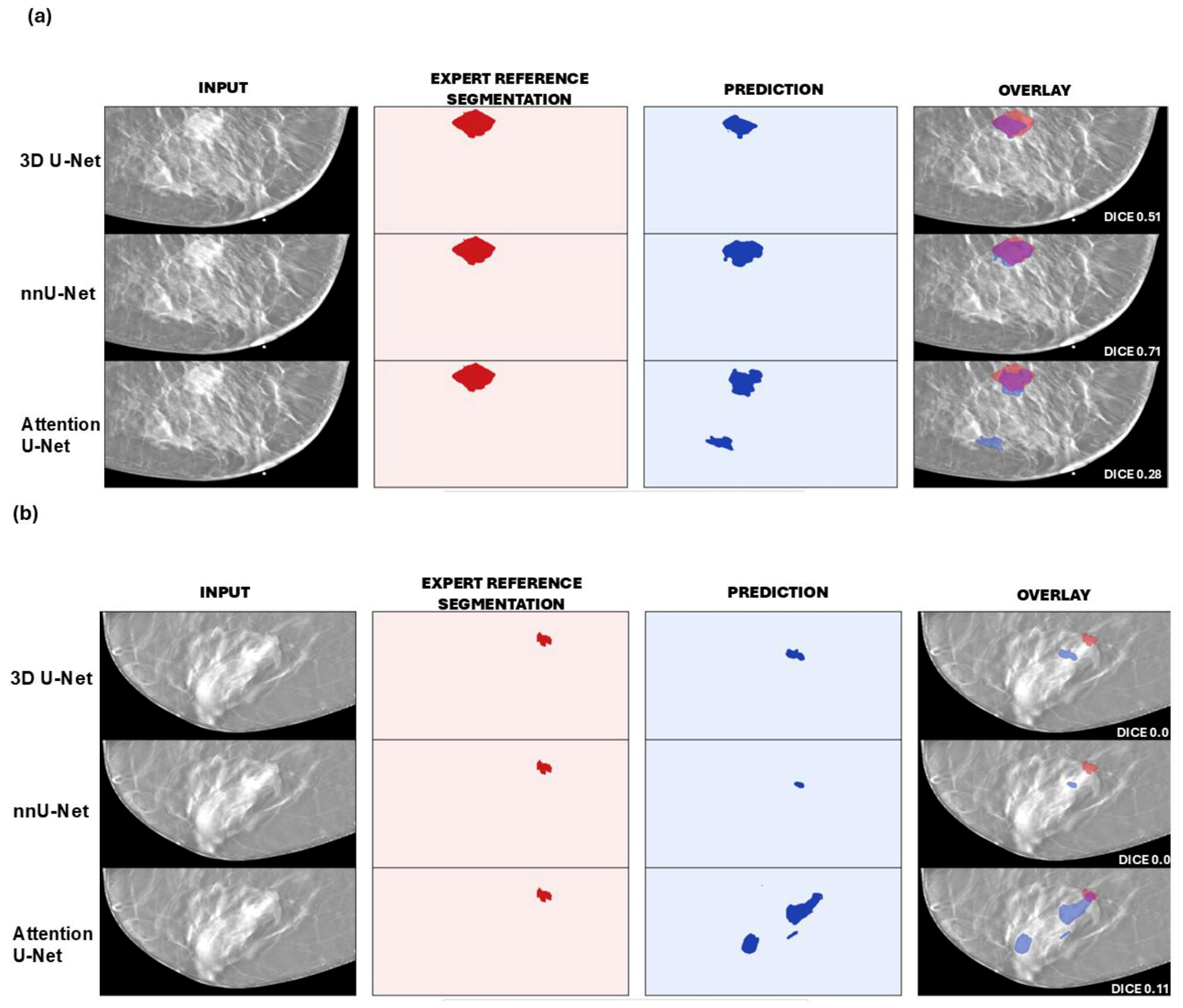
A study published in the journal Diagnostics proposes a method for AI algorithms to learn to outline breast tumors in tomosynthesis. The models are first trained on computer-simulated images and then fine-tuned with a very small number of real exams.
Digital breast tomosynthesis (DBT) is one of the most promising techniques for detecting breast cancer. Unlike conventional mammography, it offers a quasi-three-dimensional view that reduces the effect of overlapping tissue, which is especially useful in women with dense breasts. That extra information comes at a cost: radiologists must review dozens of slices per exam, which increases reading time and workload.
That is why artificial intelligence has drawn so much interest in this field. However, most developments have focused on detecting lesions rather than segmenting them, that is, precisely defining their boundaries. For Cristina Alfaro, first author of the study, iHEALTH researcher and faculty member at Universidad de Tarapacá, these are very different tasks.
"Detecting a tumor means, in simple terms, answering 'there is a lesion here.' Segmenting is much harder: the algorithm has to define exactly where the tumor begins and ends, pixel by pixel or voxel by voxel," the researcher explains. In tomosynthesis the challenge is greater because "we don't have a perfect three-dimensional image; it's a pseudo-3D modality, with limited depth resolution and a lot of overlapping breast tissue."
Having that complete outline can be very valuable. A volumetric segmentation "allows us to better characterize the lesion's spatial extent, shape and relationship with the surrounding tissue," says Cristina, and in the future it could support more reproducible and quantitative assessments. Even so, she notes that the method does not yet provide "a definitive clinical measurement of tumor volume."
The bottleneck: few annotated data
To train a segmentation system, it is not enough to know that an image contains cancer. A specialist has to carefully mark the tumor's contour on each slice, and that work is slow and costly. "It's an international problem. There are public mammography databases, but there are far fewer for tomosynthesis, and even fewer with precisely outlined tumors," Alfaro says. Added to this are patient privacy, the storage of large volumes of images, and differences between equipment and institutions. For this reason, she states, the scarcity of annotated data is today "one of the main bottlenecks of artificial intelligence applied to medical imaging."
The team, which also includes Gabriel Guerra from Universidad de Valparaíso, and Claudia Prieto and Domingo Mery, both from Pontificia Universidad Católica de Chile, sought a way forward in synthetic data: images generated in silico with VICTRE, a simulation software developed by Badano and colleagues and validated by the FDA.
"These aren't images created simply to 'look like' a breast," Cristina clarifies. "We start from digital models of breast anatomy and simulate both the presence of the tumor and elements of the tomosynthesis image-formation process." With this approach, the group developed BreasTomo-Synth, a dataset in which the exact location and characteristics of the tumor are known, making it possible to study how different algorithms respond under controlled conditions.
From simulated to clinical
The study used 120 synthetic cases with tumors of different sizes and 20 real clinical exams from the public BCS-DBT database, all with biopsy-confirmed malignant tumors. Three deep learning architectures were evaluated: 3D U-Net, nnU-Net and Attention U-Net. Unlike many previous studies, the models did not receive an image already cropped around the lesion. They worked with blocks of 15 slices that preserved the entire breast, so they had to both find the tumor and outline it.
The strategy had two stages. "We used the synthetic data so the models could initially learn the segmentation task and then adapted them with a limited number of real clinical images," Alfaro explains. Only 10 clinical exams were used for this adaptation, while the other 10 were set aside for the final evaluation with patients the system had never seen. Finally, the team combined the three architectures into an ensemble of models.
Results were measured using the Dice coefficient, which indicates how closely the algorithm's outline matches an expert's (0 means no overlap and 1 means perfect overlap). On synthetic images, nnU-Net reached 0.86 for large tumors. In real patients, performance was lower: the ensemble achieved the best average (0.52), followed by nnU-Net (0.48). The ensemble's advantage was statistically significant compared with Attention U-Net and 3D U-Net, but not compared with nnU-Net.
That drop when moving from one setting to the other is known as the domain gap, and it is one of the study's central findings. "Good performance on synthetic data does not guarantee exactly the same behavior on clinical images," the researcher cautions. "This reinforces the idea that synthetic data are a very valuable tool, but they must be complemented by real clinical validation."
Despite this gap, the team maintains that simulations are worth continuing to use. "If we waited until we had large, perfectly annotated clinical databases, many methodological questions could take years to investigate," Alfaro says. For her, synthetic data do not replace real data. They make it possible to move forward sooner and save clinical data for where they are irreplaceable: "in clinical validation," the next step.
Alfaro, C., Guerra, G., Prieto, C., & Mery, D. (2026). Synthetic-to-clinical ensemble learning for volumetric breast tumor segmentation in digital breast tomosynthesis under limited annotated data. Diagnostics, 16, 3046. https://doi.org/10.3390/diagnostics16183046
