Seminario 21 de Octubre 2025
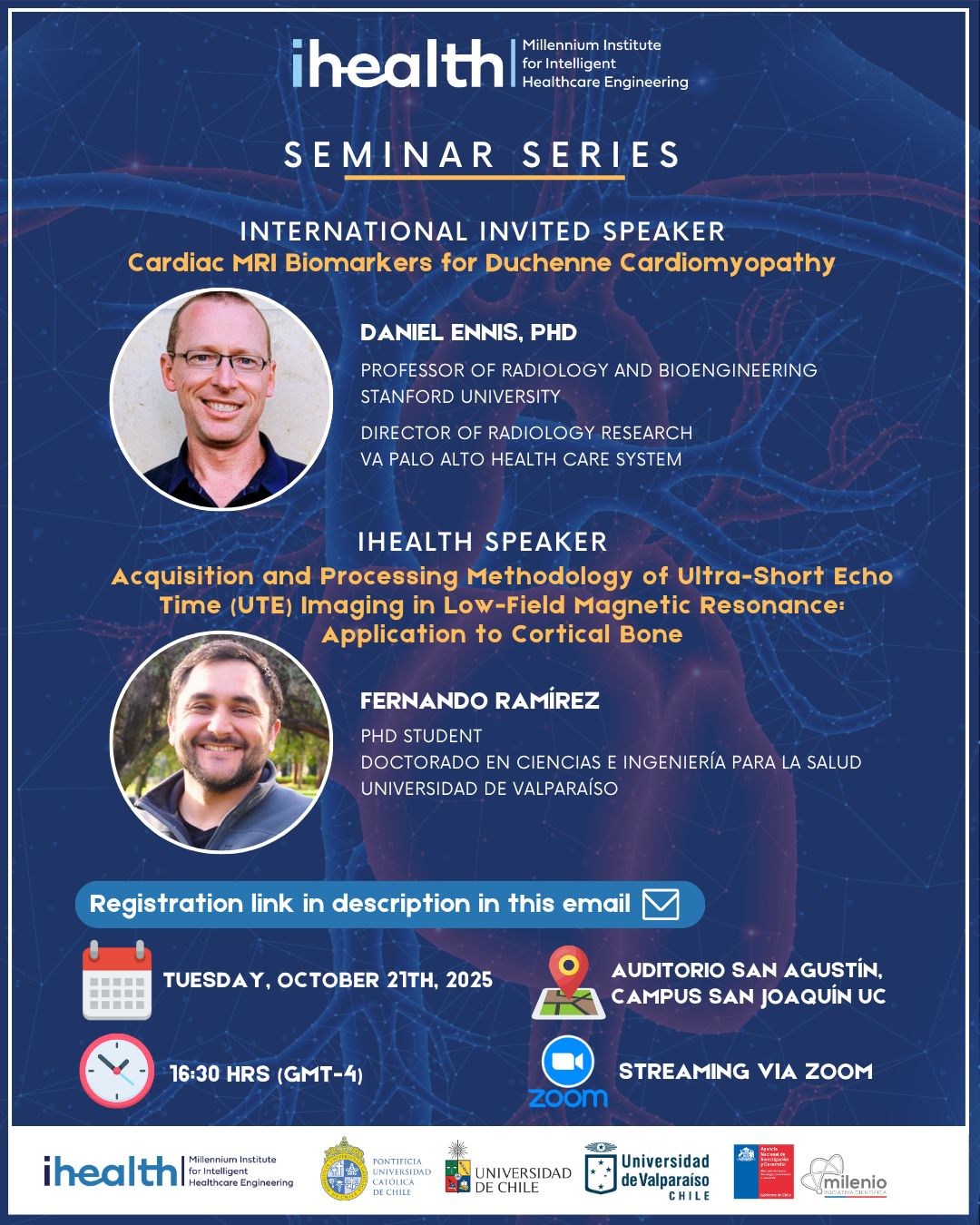
Horario: 16:30 - 18:00
INTERNATIONAL INVITED SPEAKER
Daniel Ennis
SHORT BIO: Dr. Daniel Ennis is a Professor of Radiology and Bioengineering at Stanford University. Trained at Johns Hopkins University, where he collaborated with the NIH’s Laboratory of Cardiac Energetics, he now leads the Radiological Sciences Lab and directs Radiology Research at the VA Palo Alto Health Care System. His research focuses on developing advanced cardiovascular MRI techniques to quantify cardiac structure, function, flow, and remodeling in both adults and children.
TITLE: Cardiac MRI Biomarkers for Duchenne Cardiomyopathy
ABSTRACT: Duchenne cardiomyopathy develops as a consequence of a common genetic disorder that profoundly impacts skeletal, respiratory, and cardiac muscle. The genetic variants (genotypes) are numerous; therefore, the health consequences (phenotypes) are also variable. Therefore, the specific disease course for an individual is not readily known. It is important to understand how affected (or not) a specific boy's heart may be by the disorder, but owing to their limited mobility with aging, it can be difficult to gauge heart health absent the ability to exercise. Medical imaging plays an important role in understanding the impact of the disease. Our lab has spent decades developing magnetic resonance imaging (MRI) methods to better understand heart health and disease.
iHEALTH SPEAKER
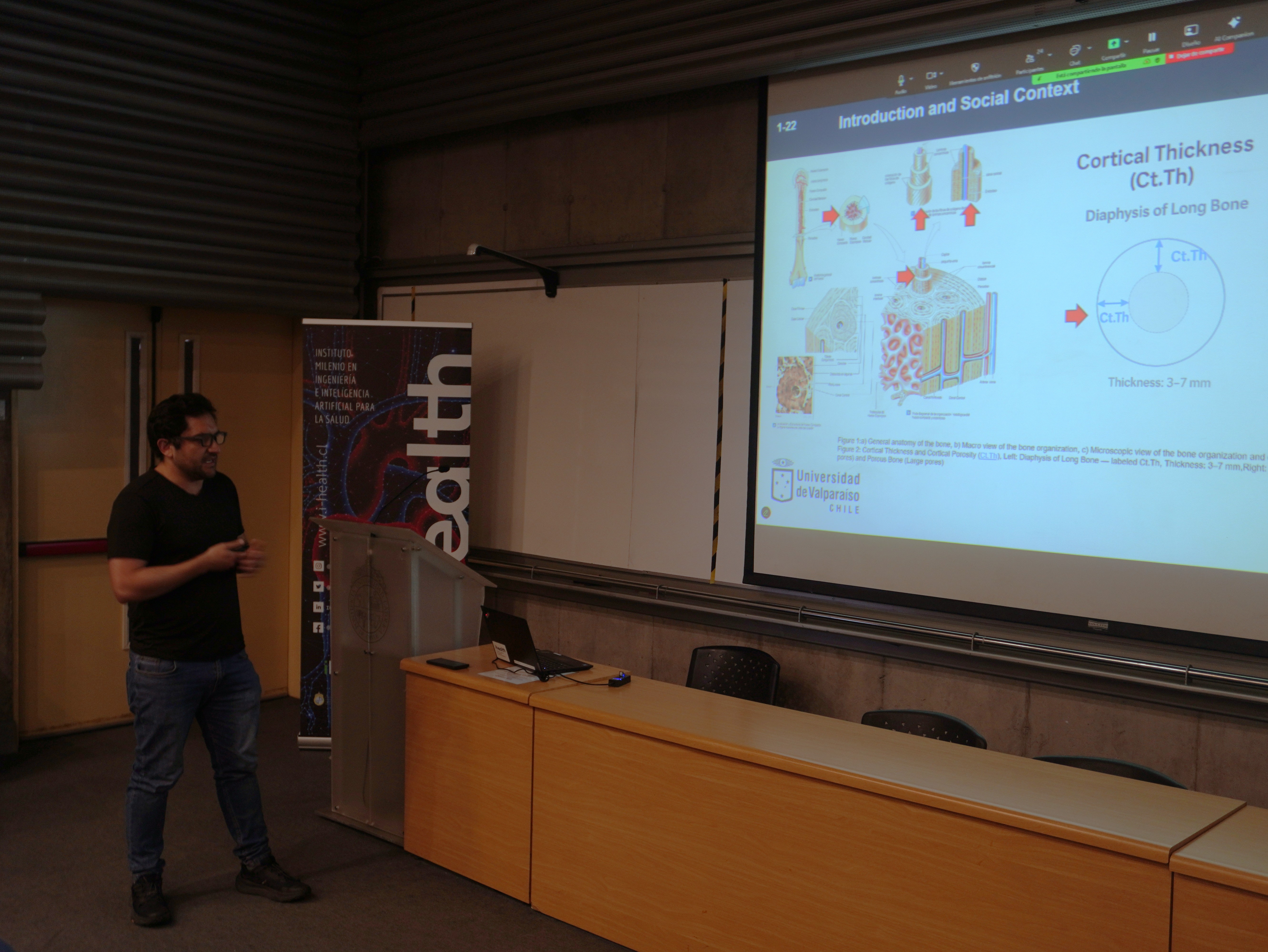
Fernando Ramírez
SHORT BIO: Fernando Ramírez is a PhD candidate in Applied Computer Engineering at the University of Valparaíso, Chile. His research focuses on the use of MRI and ultrasound (BDAT) for assessing bone fragility, integrating computational and imaging approaches to enhance diagnostic precision. He holds a degree in Biomedical Engineering and has professional experience in healthcare project management and hospital operations.
TITLE: Acquisition and Processing Methodology of Ultra-Short Echo Time (UTE) Imaging in Low-Field Magnetic Resonance: Application to Cortical Bone
ABSTRACT: Cortical bone features ultrashort T2* decay, rendering it invisible in conventional low-field MRI (0.55 T). To enable imaging, we optimized a UTE-2D pulse sequence utilizing ramp sampling and a radial trajectory to achieve minimum echo times (TE ≈ 20 μs) and incorporate necessary spoiling mechanisms. This technical adaptation addresses the inherent low signal-to-noise ratio (SNR) challenge at 0.55 T. The primary objective is to validate the methodology and quantify the MRI Porosity Index (PI), a biomarker derived from UTE signals sensitive to bone microarchitecture and fragility. Final experimental validation compares the MRI Porosity Index (PI) with established structural and mechanical indicators—Cortical Porosity (Ct.Po) and Cortical Thickness (Ct.Th)—derived from Bi-directional Axial Transmission (BDAT) Ultrasound.